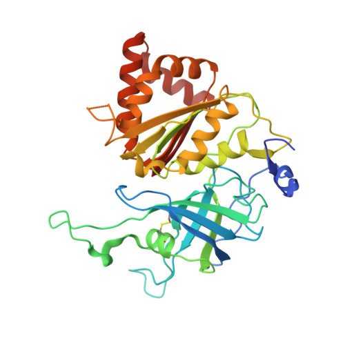
1JB9

Crystal Structure of The Ferredoxin:NADP+ Reductase From Maize Root AT 1.7 Angstroms


Experimental Data Snapshot

  • Method: X-RAY DIFFRACTION
  • Resolution: 1.70 Å
  • R-Value Free: 0.228 
  • R-Value Work: 0.164 
  • R-Value Observed: 0.167 

wwPDB Validation   3D Report Full Report


Ligand Structure Quality Assessment 


This is version 1.6 of the entry. See complete history


Literature

Biochemical and crystallographic characterization of ferredoxin-NADP(+) reductase from nonphotosynthetic tissues.

Aliverti, A.Faber, R.Finnerty, C.M.Ferioli, C.Pandini, V.Negri, A.Karplus, P.A.Zanetti, G.

(2001) Biochemistry 40: 14501-14508

  • DOI: https://doi.org/10.1021/bi011224c
  • Primary Citation of Related Structures:  
    1JB9, 3LVB

  • PubMed Abstract: 

    Distinct forms of ferredoxin-NADP(+) reductase are expressed in photosynthetic and nonphotosynthetic plant tissues. Both enzymes catalyze electron transfer between NADP(H) and ferredoxin; whereas in leaves the enzyme transfers reducing equivalents from photoreduced ferredoxin to NADP(+) in photosynthesis, in roots it has the opposite physiological role, reducing ferredoxin at the expense of NADPH mainly for use in nitrate assimilation. Here, structural and kinetic properties of a nonphotosynthetic isoform were analyzed to define characteristics that may be related to tissue-specific function. Compared with spinach leaf ferredoxin-NADP(+) reductase, the recombinant corn root isoform showed a slightly altered absorption spectrum, a higher pI, a >30-fold higher affinity for NADP(+), greater susceptibility to limited proteolysis, and an approximately 20 mV more positive redox potential. The 1.7 A resolution crystal structure is very similar to the structures of ferredoxin-NADP(+) reductases from photosynthetic tissues. Four distinct structural features of this root ferredoxin-NADP(+) reductases are an alternate conformation of the bound FAD molecule, an alternate path for the amino-terminal extension, a disulfide bond in the FAD-binding domain, and changes in the surface that binds ferredoxin.


  • Organizational Affiliation

    Dipartimento di Fisiologia e Biochimica Generali, Università degli Studi di Milano, Via Celoria 26, 20133 Milano, Italy.


Macromolecules
Find similar proteins by:  (by identity cutoff)  |  3D Structure
Entity ID: 1
MoleculeChains Sequence LengthOrganismDetailsImage
ferredoxin-NADP reductase316Zea maysMutation(s): 0 
EC: 1.18.1.2
UniProt
Find proteins for Q41736 (Zea mays)
Explore Q41736 
Go to UniProtKB:  Q41736
Entity Groups  
Sequence Clusters30% Identity50% Identity70% Identity90% Identity95% Identity100% Identity
UniProt GroupQ41736
Sequence Annotations
Expand
  • Reference Sequence
Small Molecules
Ligands 1 Unique
IDChains Name / Formula / InChI Key2D Diagram3D Interactions
FAD
Query on FAD

Download Ideal Coordinates CCD File 
B [auth A]FLAVIN-ADENINE DINUCLEOTIDE
C27 H33 N9 O15 P2
VWWQXMAJTJZDQX-UYBVJOGSSA-N
Experimental Data & Validation

Experimental Data

  • Method: X-RAY DIFFRACTION
  • Resolution: 1.70 Å
  • R-Value Free: 0.228 
  • R-Value Work: 0.164 
  • R-Value Observed: 0.167 
  • Space Group: P 32 2 1
Unit Cell:
Length ( Å )Angle ( ˚ )
a = 59.67α = 90
b = 59.67β = 90
c = 189.1γ = 120
Software Package:
Software NamePurpose
SCALEPACKdata scaling
MRXmodel building
TNTrefinement
MRXphasing

Structure Validation

View Full Validation Report


Ligand Structure Quality Assessment 


Entry History 

Deposition Data

Revision History  (Full details and data files)

  • Version 1.0: 2001-07-04
    Type: Initial release
  • Version 1.1: 2008-04-27
    Changes: Version format compliance
  • Version 1.2: 2011-07-13
    Changes: Version format compliance
  • Version 1.3: 2012-07-11
    Changes: Database references
  • Version 1.4: 2014-03-26
    Changes: Database references
  • Version 1.5: 2017-10-04
    Changes: Refinement description
  • Version 1.6: 2023-08-16
    Changes: Data collection, Database references, Derived calculations, Refinement description