Phospholipase A2 engineering. Deletion of the C-terminus segment changes substrate specificity and uncouples calcium and substrate binding at the zwitterionic interface.
Huang, B., Yu, B.Z., Rogers, J., Byeon, I.J., Sekar, K., Chen, X., Sundaralingam, M., Tsai, M.D., Jain, M.K.(1996) Biochemistry 35: 12164-12174
- PubMed: 8810924
- DOI: https://doi.org/10.1021/bi960234o
- Primary Citation of Related Structures:
1IRB - PubMed Abstract:
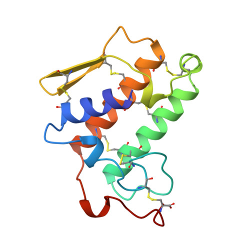
It has been suggested [Dijkstra, B. W., Drenth, J., & Kalk, K. H. (1981) Nature 289, 604-606] that the interfacial binding site of phospholipase A2 (PLA2) involves a large number of residues, including a cluster at the N-terminus and another cluster at the C-terminus. The approaches of multiple mutation and deletion were used to test the roles of the C-terminal residues of bovine pancreatic PLA2 overexpressed in Escherichia coli. A double mutant K120A/K121A and a deletion mutant delta 115-123/ C27A were constructed, and structural and functional analyses were performed on both mutants. The double mutant showed little perturbation in the global structure on the basis of proton NMR and X-ray crystallographic analyses. The proton NMR analysis of the deletion mutant suggested that a few residues at the active site, the hydrophobic channel, and the calcium binding loop are perturbed, but the global conformation is not changed. The mutants were then characterized for catalytic and binding properties by use of various kinetic and spectroscopic methods. The double mutant behaved in a manner similar to that of the wild type (WT) PLA2 in every property examined. The deletion mutant was found to show an interesting change of substrate specificity. The kcat,app of the zwitterionic DC8PC micelles but not the anionic DC8PM micelles decreased by a factor of > 100; however, the activity of DC8PC was restored upon addition of 4 M NaCl. The results of fluorescence spectroscopic studies indicate that the deletion mutant behaves in a manner similar to that of WT in the binding to anionic vesicles and to zwitterionic neutral diluent. Thus, the binding affinity of the enzyme to the interface (the E to E* step) should not be the main cause for the change in substrate specificity. The cause lies at least partially in the binding of substrate or inhibitor to the active site of the enzyme at the interface, i.e., the E* to E*L step, as revealed by the results of equilibrium binding studies. The equilibrium dissociation constants of ligands are generally higher for the deletion mutant (relative to WT) at the zwitterionic interface but not at the anionic interface. The cause for the low affinity of an active site-directed ligand to the active site at the zwitterionic interface could be related to the inability of Ca2+ to enhance ligand binding for the deletion mutant. This is in contrast to the WT PLA2 for which Ca2+ binding enhances binding of the substrate to the active site. Overall, the structural and functional perturbations caused by deleting the C-terminal segment are modest, but the changes in substrate specificity and the uncoupling between substrate and calcium binding are interesting and significant.
Organizational Affiliation:
Department of Chemistry, Ohio State University, Columbus 43210, USA.