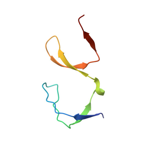
Effect of pH and salt bridges on structural assembly: molecular structures of the monomer and intertwined dimer of the Eps8 SH3 domain.
Kishan, K.V., Newcomer, M.E., Rhodes, T.H., Guilliot, S.D.(2001) Protein Sci 10: 1046-1055
- PubMed: 11316885
- DOI: https://doi.org/10.1110/ps.50401
- Primary Citation of Related Structures:
1I07, 1I0C - PubMed Abstract:
The SH3 domain of Eps8 was previously found to form an intertwined, domain-swapped dimer. We report here a monomeric structure of the EPS8 SH3 domain obtained from crystals grown at low pH, as well as an improved domain-swapped dimer structure at 1.8 A resolution. In the domain-swapped dimer the asymmetric unit contains two "hybrid-monomers." In the low pH form there are two independently folded SH3 molecules per asymmetric unit. The formation of intermolecular salt bridges is thought to be the reason for the formation of the dimer. On the basis of the monomer SH3 structure, it is argued that Eps8 SH3 should, in principle, bind to peptides containing a PxxP motif. Recently it was reported that Eps8 SH3 binds to a peptide with a PxxDY motif. Because the "SH3 fold" is conserved, alternate binding sites may be possible for the PxxDY motif to bind. The strand exchange or domain swap occurs at the n-src loops because the n-src loops are flexible. The thermal b-factors also indicate the flexible nature of n-src loops and a possible handle for domain swap initiation. Despite the loop swapping, the typical SH3 fold in both forms is conserved structurally. The interface of the acidic form of SH3 is stabilized by a tetragonal network of water molecules above hydrophobic residues. The intertwined dimer interface is stabilized by hydrophobic and aromatic stacking interactions in the core and by hydrophilic interactions on the surface.
Organizational Affiliation:
Institute of Microbial Technology, Sector 39-A, Chandigarh 160 036, India. kishan@imtech.res.in