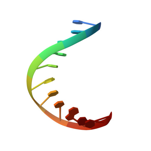
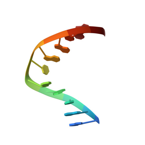
Solution structure of the 2-amino-1- methyl-6-phenylimidazo[4,5-b]pyridine C8-deoxyguanosine adduct in duplex DNA.
Brown, K., Hingerty, B.E., Guenther, E.A., Krishnan, V.V., Broyde, S., Turteltaub, K.W., Cosman, M.(2001) Proc Natl Acad Sci U S A 98: 8507-8512
- PubMed: 11438709
- DOI: https://doi.org/10.1073/pnas.151251898
- Primary Citation of Related Structures:
1HZ0 - PubMed Abstract:
The carcinogenic heterocyclic amine (HA) 2-amino-1-methyl-6-phenylimidazo[4,5-b]pyridine (PhIP) is formed during the cooking of various meats. To enable structure/activity studies aimed at understanding how DNA damaged by a member of the HA class of compounds can ultimately lead to cancer, we have determined the first solution structure of an 11-mer duplex containing the C8-dG adduct formed by reaction with N-acetoxy-PhIP. A slow conformational exchange is observed in which the PhIP ligand either intercalates into the DNA helix by denaturing and displacing the modified base pair (main form) or is located outside the helix in a minimally perturbed B-DNA duplex (minor form). In the main base-displaced intercalation structure, the minor groove is widened, and the major groove is compressed at the lesion site because of the location of the bulky PhIP-N-methyl and phenyl ring in the minor groove; this distortion causes significant bending of the helix. The PhIP phenyl ring interacts with the phosphodiester-sugar ring backbone of the complementary strand and its fast rotation with respect to the intercalated imidazopyridine ring causes substantial distortions at this site, such as unwinding and bulging-out of the strand. The glycosidic torsion angle of the [PhIP]dG residue is syn, and the displaced guanine base is directed toward the 3' end of the modified strand. This study contributes, to our knowledge, the first structural information on the biologically relevant HA class to a growing body of knowledge about how conformational similarities and differences for a variety of types of lesions can influence protein interactions and ultimately biological outcome.
Organizational Affiliation:
Biology and Biotechnology Research Program, Lawrence Livermore National Laboratory, Livermore, CA 94551, USA.