1HE9

Crystal structure of the GAP domain of the Pseudomonas aeruginosa ExoS toxin


Experimental Data Snapshot

  • Method: X-RAY DIFFRACTION
  • Resolution: 2.40 Å
  • R-Value Free: 0.265 
  • R-Value Work: 0.257 
  • R-Value Observed: 0.257 

wwPDB Validation   3D Report Full Report


This is version 1.2 of the entry. See complete history


Literature

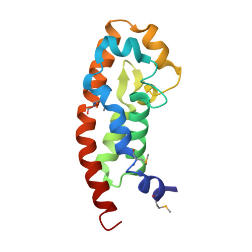
Structure of the Exos Gtpase Activating Domain

Wurtele, M.Renault, L.Barbieri, J.T.Wittinghofer, A.Wolf, E.

(2001) FEBS Lett 491: 26

  • DOI: https://doi.org/10.1016/s0014-5793(01)02105-6
  • Primary Citation of Related Structures:  
    1HE9

  • PubMed Abstract: 

    Pseudomonas aeruginosa is an opportunistic bacterial pathogen of great medical relevance. One of its major toxins, exoenzyme S (ExoS), is a dual function protein with a C-terminal Ras-ADP-ribosylation domain and an N-terminal GTPase activating protein (GAP) domain specific for Rho-family proteins. We report here the three-dimensional structure of the N-terminal domain of ExoS determined by X-ray crystallography to 2.4 A resolution. Its fold is all helical with a four helix bundle core capped by additional irregular helices. Loops that are known to interact with Rho-family proteins show very large mobility. Considering the importance of ExoS in Pseudomonas pathogenicity, this structure could be of interest for drug targeting.


  • Organizational Affiliation

    Max-Planck-Institut für molekulare Physiologie, Abteilung Strukturelle Biologie, Otto-Hahn-Str. 11, 44227 Dortmund, Germany.


Macromolecules
Find similar proteins by:  (by identity cutoff)  |  3D Structure
Entity ID: 1
MoleculeChains Sequence LengthOrganismDetailsImage
EXOENZYME S134Pseudomonas aeruginosaMutation(s): 3 
Gene Names: EXOS
UniProt
Find proteins for Q51451 (Pseudomonas aeruginosa)
Explore Q51451 
Go to UniProtKB:  Q51451
Entity Groups  
Sequence Clusters30% Identity50% Identity70% Identity90% Identity95% Identity100% Identity
UniProt GroupQ51451
Sequence Annotations
Expand
  • Reference Sequence
Small Molecules
Modified Residues  1 Unique
IDChains TypeFormula2D DiagramParent
MSE
Query on MSE
A
L-PEPTIDE LINKINGC5 H11 N O2 SeMET
Experimental Data & Validation

Experimental Data

  • Method: X-RAY DIFFRACTION
  • Resolution: 2.40 Å
  • R-Value Free: 0.265 
  • R-Value Work: 0.257 
  • R-Value Observed: 0.257 
  • Space Group: C 2 2 21
Unit Cell:
Length ( Å )Angle ( ˚ )
a = 42.897α = 90
b = 55.646β = 90
c = 107.472γ = 90
Software Package:
Software NamePurpose
CNSrefinement
DENZOdata reduction
SCALEPACKdata scaling
SHARPphasing

Structure Validation

View Full Validation Report


Entry History 

Deposition Data

Revision History  (Full details and data files)

  • Version 1.0: 2001-03-19
    Type: Initial release
  • Version 1.1: 2011-05-08
    Changes: Version format compliance
  • Version 1.2: 2011-07-13
    Changes: Version format compliance