1HCU

alpha-1,2-mannosidase from Trichoderma reesei


Experimental Data Snapshot

  • Method: X-RAY DIFFRACTION
  • Resolution: 2.37 Å
  • R-Value Free: 0.232 
  • R-Value Work: 0.177 
  • R-Value Observed: 0.177 

wwPDB Validation   3D Report Full Report


Ligand Structure Quality Assessment 


This is version 1.6 of the entry. See complete history


Literature

Trichoderma Reesei Alpha-1,2-Mannosidase: Structural Basis for the Cleavage of Four Consecutive Mannose Residues

Van Petegem, F.Contreras, H.Contreras, R.Van Beeumen, J.

(2001) J Mol Biol 312: 157

  • DOI: https://doi.org/10.1006/jmbi.2001.4946
  • Primary Citation of Related Structures:  
    1HCU

  • PubMed Abstract: 

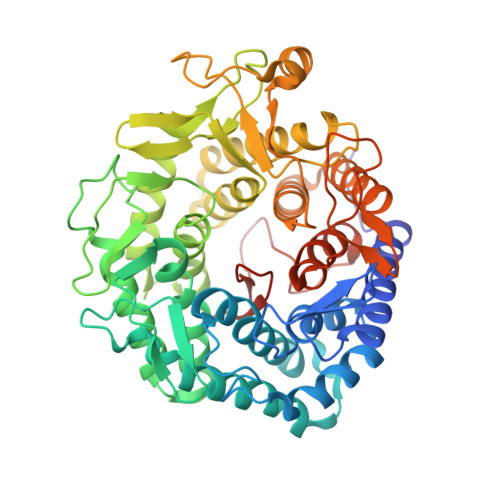
    The process of N-glycosylation of eukaryotic proteins involves a range of host enzymes that delete or add saccharide monomers. While endoplasmic reticulum (E.R.) mannosidases cleave only one mannose to produce the Man8B isomer, an alpha-1,2-mannosidase from Trichoderma reesei can sequentially cleave all four 1,2-linked mannose sugars from a Man(9)GlcNAc(2) oligosaccharide, a feature reminiscent of the activity of Golgi mannosidases. We now report the structure of the T. reesei enzyme at 2.37 A resolution. The enzyme folds as an (alpha alpha)(7) barrel. The substrate-binding site of the T. reesei mannosidase differs appreciably from the Saccharomyces cerevisiae enzyme. In the former, shorter loops at the surface allow substrate protein to come closer to the catalytic site. There is more internal space available, so that different oligosaccharide conformations are sterically allowed in the T. reesei alpha-1,2-mannosidase.


  • Organizational Affiliation

    Department of Biochemistry, Physiology and Microbiology, University of Ghent, K.L. Ledeganckstraat 35, 9000 Ghent, Belgium.


Macromolecules
Find similar proteins by:  (by identity cutoff)  |  3D Structure
Entity ID: 1
MoleculeChains Sequence LengthOrganismDetailsImage
ALPHA-1,2-MANNOSIDASE
A, B, C, D
503Trichoderma reeseiMutation(s): 0 
EC: 3.2.1.113
UniProt
Find proteins for Q9P8T8 (Hypocrea jecorina)
Explore Q9P8T8 
Go to UniProtKB:  Q9P8T8
Entity Groups  
Sequence Clusters30% Identity50% Identity70% Identity90% Identity95% Identity100% Identity
UniProt GroupQ9P8T8
Sequence Annotations
Expand
  • Reference Sequence
Small Molecules
Ligands 2 Unique
IDChains Name / Formula / InChI Key2D Diagram3D Interactions
NAG
Query on NAG

Download Ideal Coordinates CCD File 
E [auth A]
F [auth A]
G [auth A]
I [auth B]
J [auth B]
E [auth A],
F [auth A],
G [auth A],
I [auth B],
J [auth B],
K [auth B],
M [auth C],
N [auth C],
P [auth D],
Q [auth D]
2-acetamido-2-deoxy-beta-D-glucopyranose
C8 H15 N O6
OVRNDRQMDRJTHS-FMDGEEDCSA-N
CA
Query on CA

Download Ideal Coordinates CCD File 
H [auth A],
L [auth B],
O [auth C],
R [auth D]
CALCIUM ION
Ca
BHPQYMZQTOCNFJ-UHFFFAOYSA-N
Experimental Data & Validation

Experimental Data

  • Method: X-RAY DIFFRACTION
  • Resolution: 2.37 Å
  • R-Value Free: 0.232 
  • R-Value Work: 0.177 
  • R-Value Observed: 0.177 
  • Space Group: P 1 21 1
Unit Cell:
Length ( Å )Angle ( ˚ )
a = 86.201α = 90
b = 106.836β = 99.42
c = 101.388γ = 90
Software Package:
Software NamePurpose
CNSrefinement
DENZOdata reduction
SCALEPACKdata scaling
AMoREphasing

Structure Validation

View Full Validation Report


Ligand Structure Quality Assessment 


Entry History 

Deposition Data

Revision History  (Full details and data files)

  • Version 1.0: 2001-10-18
    Type: Initial release
  • Version 1.1: 2011-05-08
    Changes: Version format compliance
  • Version 1.2: 2011-07-13
    Changes: Version format compliance
  • Version 1.3: 2019-05-08
    Changes: Data collection, Derived calculations, Experimental preparation, Other
  • Version 1.4: 2019-07-24
    Changes: Data collection
  • Version 1.5: 2020-07-29
    Type: Remediation
    Reason: Carbohydrate remediation
    Changes: Data collection, Derived calculations, Other, Structure summary
  • Version 1.6: 2023-12-13
    Changes: Data collection, Database references, Refinement description, Structure summary