1H8L

Duck Carboxypeptidase D Domain II in complex with GEMSA


Experimental Data Snapshot

  • Method: X-RAY DIFFRACTION
  • Resolution: 2.60 Å
  • R-Value Free: 0.239 
  • R-Value Work: 0.199 
  • R-Value Observed: 0.199 

wwPDB Validation   3D Report Full Report


This is version 3.1 of the entry. See complete history


Literature

The crystal structure of the inhibitor-complexed carboxypeptidase D domain II and the modeling of regulatory carboxypeptidases.

Aloy, P.Companys, V.Vendrell, J.Aviles, F.X.Fricker, L.D.Coll, M.Gomis-Ruth, F.X.

(2001) J Biol Chem 276: 16177-16184

  • DOI: https://doi.org/10.1074/jbc.M011457200
  • Primary Citation of Related Structures:  
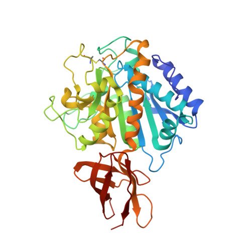
    1H8L

  • PubMed Abstract: 

    The three-dimensional crystal structure of duck carboxypeptidase D domain II has been solved in a complex with the peptidomimetic inhibitor, guanidinoethylmercaptosuccinic acid, occupying the specificity pocket. This structure allows a clear definition of the substrate binding sites and the substrate funnel-like access. The structure of domain II is the only one available from the regulatory carboxypeptidase family and can be used as a general template for its members. Here, it has been used to model the structures of domains I and III from the former protein and of human carboxypeptidase E. The models obtained show that the overall topology is similar in all cases, the main differences being local and because of insertions in non-regular loops. In both carboxypeptidase D domain I and carboxypeptidase E slightly different shapes of the access to the active site are predicted, implying some kind of structural selection of protein or peptide substrates. Furthermore, emplacement of the inhibitor structure in the active site of the constructed models showed that the inhibitor fits very well in all of them and that the relevant interactions observed with domain II are conserved in domain I and carboxypeptidase E but not in the non-active domain III because of the absence of catalytically indispensable residues in the latter protein. However, in domain III some of the residues potentially involved in substrate binding are well preserved, together with others of unknown roles, which also are highly conserved among all carboxypeptidases. These observations, taken together with others, suggest that domain III might play a role in the binding and presentation of proteins or peptide substrates, such as the pre-S domain of the large envelope protein of duck hepatitis B virus.


  • Organizational Affiliation

    Institut de Biologia Fonamental and Departament de Bioquimica i Biologia Molecular, Unitat de Ciències, Universitat Autònoma de Barcelona, 08193 Bellaterra, Barcelona, Spain.


Macromolecules
Find similar proteins by:  (by identity cutoff)  |  3D Structure
Entity ID: 1
MoleculeChains Sequence LengthOrganismDetailsImage
CARBOXYPEPTIDASE GP180 RESIDUES 503-882380Lophonetta specularioidesMutation(s): 0 
UniProt
Find proteins for P83852 (Lophonetta specularioides)
Explore P83852 
Go to UniProtKB:  P83852
Entity Groups  
Sequence Clusters30% Identity50% Identity70% Identity90% Identity95% Identity100% Identity
UniProt GroupP83852
Sequence Annotations
Expand
  • Reference Sequence
Oligosaccharides

Help

Entity ID: 2
MoleculeChains Length2D Diagram Glycosylation3D Interactions
beta-D-mannopyranose-(1-4)-2-acetamido-2-deoxy-beta-D-glucopyranose-(1-4)-2-acetamido-2-deoxy-beta-D-glucopyranose
B
3N-Glycosylation
Glycosylation Resources
GlyTouCan:  G15407YE
GlyCosmos:  G15407YE
GlyGen:  G15407YE
Entity ID: 3
MoleculeChains Length2D Diagram Glycosylation3D Interactions
2-acetamido-2-deoxy-beta-D-glucopyranose-(1-4)-2-acetamido-2-deoxy-beta-D-glucopyranose
C
2N-Glycosylation
Glycosylation Resources
GlyTouCan:  G42666HT
GlyCosmos:  G42666HT
GlyGen:  G42666HT
Small Molecules
Ligands 4 Unique
IDChains Name / Formula / InChI Key2D Diagram3D Interactions
GEM
Query on GEM

Download Ideal Coordinates CCD File 
D [auth A](2-GUANIDINOETHYLMERCAPTO)SUCCINIC ACID
C7 H13 N3 O4 S
VKVCLXDFOQQABP-BYPYZUCNSA-N
NAG
Query on NAG

Download Ideal Coordinates CCD File 
E [auth A]2-acetamido-2-deoxy-beta-D-glucopyranose
C8 H15 N O6
OVRNDRQMDRJTHS-FMDGEEDCSA-N
SO4
Query on SO4

Download Ideal Coordinates CCD File 
F [auth A]SULFATE ION
O4 S
QAOWNCQODCNURD-UHFFFAOYSA-L
ZN
Query on ZN

Download Ideal Coordinates CCD File 
G [auth A]ZINC ION
Zn
PTFCDOFLOPIGGS-UHFFFAOYSA-N
Binding Affinity Annotations 
IDSourceBinding Affinity
GEM PDBBind:  1H8L Ki: 34 (nM) from 1 assay(s)
Experimental Data & Validation

Experimental Data

  • Method: X-RAY DIFFRACTION
  • Resolution: 2.60 Å
  • R-Value Free: 0.239 
  • R-Value Work: 0.199 
  • R-Value Observed: 0.199 
  • Space Group: P 21 3
Unit Cell:
Length ( Å )Angle ( ˚ )
a = 136.48α = 90
b = 136.48β = 90
c = 136.48γ = 90
Software Package:
Software NamePurpose
CNSrefinement
MOSFLMdata reduction
SCALAdata scaling

Structure Validation

View Full Validation Report


Entry History 

Deposition Data

Revision History  (Full details and data files)

  • Version 1.0: 2002-02-08
    Type: Initial release
  • Version 1.1: 2011-05-07
    Changes: Version format compliance
  • Version 1.2: 2011-07-13
    Changes: Version format compliance
  • Version 2.0: 2018-07-11
    Changes: Advisory, Atomic model, Data collection, Database references, Derived calculations, Non-polymer description, Source and taxonomy, Structure summary
  • Version 2.1: 2019-07-10
    Changes: Data collection, Derived calculations
  • Version 2.2: 2019-07-24
    Changes: Data collection
  • Version 3.0: 2020-07-29
    Type: Remediation
    Reason: Carbohydrate remediation
    Changes: Advisory, Atomic model, Data collection, Derived calculations, Structure summary
  • Version 3.1: 2023-12-13
    Changes: Data collection, Database references, Refinement description, Structure summary