1H4J

Methylobacterium extorquens methanol dehydrogenase D303E mutant


Experimental Data Snapshot

  • Method: X-RAY DIFFRACTION
  • Resolution: 3.00 Å
  • R-Value Free: 0.217 
  • R-Value Work: 0.187 
  • R-Value Observed: 0.187 

wwPDB Validation   3D Report Full Report


Ligand Structure Quality Assessment 


This is version 1.3 of the entry. See complete history


Literature

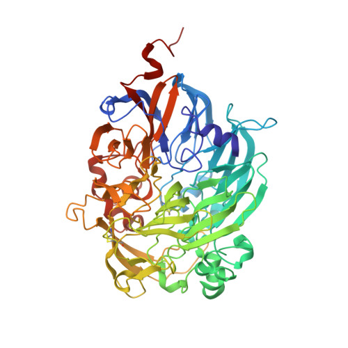
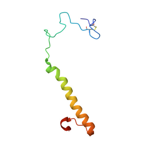
Site-Directed Mutagenesis and X-Ray Crystallography of the Pqq-Containing Quinoprotein Methanol Dehydrogenase and its Electron Acceptor, Cytochrome C(L)(,)

Afolabi, P.R.Mohammed, F.Amaratunga, K.Majekodunmi, O.Dales, S.L.Gill, R.Thompson, D.Cooper, J.B.Wood, S.P.Goodwin, P.M.Anthony, C.

(2001) Biochemistry 40: 9799-9809

  • DOI: https://doi.org/10.1021/bi002932l
  • Primary Citation of Related Structures:  
    1H4J

  • PubMed Abstract: 

    Two proteins specifically involved in methanol oxidation in the methylotrophic bacterium Methylobacterium extorquens have been modified by site-directed mutagenesis. Mutation of the proposed active site base (Asp303) to glutamate in methanol dehydrogenase (MDH) gave an active enzyme (D303E-MDH) with a greatly reduced affinity for substrate and with a lower activation energy. Results of kinetic and deuterium isotope studies showed that the essential mechanism in the mutant protein was unchanged, and that the step requiring activation by ammonia remained rate limiting. No spectrally detectable intermediates could be observed during the reaction. The X-ray structure, determined to 3 A resolution, of D303E-MDH showed that the position and coordination geometry of the Ca2+ ion in the active site was altered; the larger Glu303 side chain was coordinated to the Ca2+ ion and also hydrogen bonded to the O5 atom of pyrroloquinoline quinone (PQQ). The properties and structure of the D303E-MDH are consistent with the previous proposal that the reaction in MDH is initiated by proton abstraction involving Asp303, and that the mechanism involves a direct hydride transfer reaction. Mutation of the two adjacent cysteine residues that make up the novel disulfide ring in the active site of MDH led to an inactive enzyme, confirming the essential role of this remarkable ring structure. Mutations of cytochrome c(L), which is the electron acceptor from MDH was used to identify Met109 as the sixth ligand to the heme.


  • Organizational Affiliation

    Division of Biochemistry and Molecular Biology, School of Biological Sciences, University of Southampton, Southampton SO16 7PX, UK.


Macromolecules
Find similar proteins by:  (by identity cutoff)  |  3D Structure
Entity ID: 1
MoleculeChains Sequence LengthOrganismDetailsImage
Methanol dehydrogenase [cytochrome c] subunit 1
A, C, E, G
599Methylorubrum extorquensMutation(s): 1 
Gene Names: moxFmxaFMexAM1_META1p4538
EC: 1.1.2.7
UniProt
Find proteins for P16027 (Methylorubrum extorquens (strain ATCC 14718 / DSM 1338 / JCM 2805 / NCIMB 9133 / AM1))
Explore P16027 
Go to UniProtKB:  P16027
Entity Groups  
Sequence Clusters30% Identity50% Identity70% Identity90% Identity95% Identity100% Identity
UniProt GroupP16027
Sequence Annotations
Expand
  • Reference Sequence
Find similar proteins by:  (by identity cutoff)  |  3D Structure
Entity ID: 2
MoleculeChains Sequence LengthOrganismDetailsImage
Methanol dehydrogenase [cytochrome c] subunit 2
B, D, F, H
74Methylorubrum extorquensMutation(s): 0 
Gene Names: moxImxaIMexAM1_META1p4535
EC: 1.1.2.7
UniProt
Find proteins for P14775 (Methylorubrum extorquens (strain ATCC 14718 / DSM 1338 / JCM 2805 / NCIMB 9133 / AM1))
Explore P14775 
Go to UniProtKB:  P14775
Entity Groups  
Sequence Clusters30% Identity50% Identity70% Identity90% Identity95% Identity100% Identity
UniProt GroupP14775
Sequence Annotations
Expand
  • Reference Sequence
Experimental Data & Validation

Experimental Data

  • Method: X-RAY DIFFRACTION
  • Resolution: 3.00 Å
  • R-Value Free: 0.217 
  • R-Value Work: 0.187 
  • R-Value Observed: 0.187 
  • Space Group: P 1 21 1
Unit Cell:
Length ( Å )Angle ( ˚ )
a = 101.85α = 90
b = 61.02β = 92.83
c = 212.68γ = 90
Software Package:
Software NamePurpose
X-PLORrefinement
MOSFLMdata reduction
CCP4data scaling
MOLREPphasing

Structure Validation

View Full Validation Report


Ligand Structure Quality Assessment 


Entry History 

Deposition Data

Revision History  (Full details and data files)

  • Version 1.0: 2001-08-23
    Type: Initial release
  • Version 1.1: 2011-05-08
    Changes: Version format compliance
  • Version 1.2: 2011-07-13
    Changes: Version format compliance
  • Version 1.3: 2018-09-19
    Changes: Advisory, Data collection, Database references, Source and taxonomy, Structure summary