1H2S

Molecular basis of transmenbrane signalling by sensory rhodopsin II-transducer complex


Experimental Data Snapshot

  • Method: X-RAY DIFFRACTION
  • Resolution: 1.93 Å
  • R-Value Free: 0.258 
  • R-Value Work: 0.226 
  • R-Value Observed: 0.226 

wwPDB Validation   3D Report Full Report


This is version 1.6 of the entry. See complete history


Literature

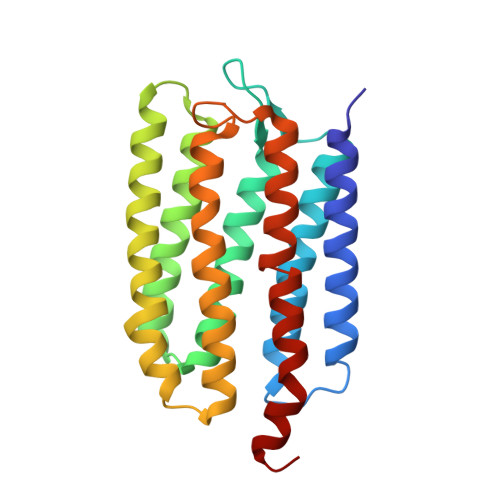
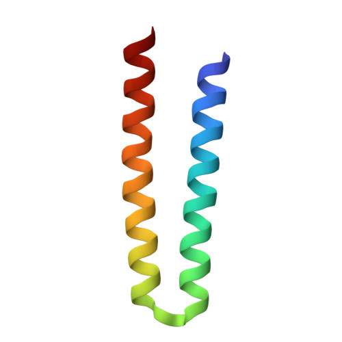
Molecular Basis of Transmembrane Signalling by Sensory Rhodopsin II-Transducer Complex

Gordeliy, V.I.Labahn, J.Moukhametzianov, R.Efremov, R.Granzin, J.Schlesinger, R.Bueldt, G.Savopol, T.Scheidig, A.Klare, J.P.Engelhard, M.

(2002) Nature 419: 484

  • DOI: https://doi.org/10.1038/nature01109
  • Primary Citation of Related Structures:  
    1H2S

  • PubMed Abstract: 

    Microbial rhodopsins, which constitute a family of seven-helix membrane proteins with retinal as a prosthetic group, are distributed throughout the Bacteria, Archaea and Eukaryota. This family of photoactive proteins uses a common structural design for two distinct functions: light-driven ion transport and phototaxis. The sensors activate a signal transduction chain similar to that of the two-component system of eubacterial chemotaxis. The link between the photoreceptor and the following cytoplasmic signal cascade is formed by a transducer molecule that binds tightly and specifically to its cognate receptor by means of two transmembrane helices (TM1 and TM2). It is thought that light excitation of sensory rhodopsin II from Natronobacterium pharaonis (SRII) in complex with its transducer (HtrII) induces an outward movement of its helix F (ref. 6), which in turn triggers a rotation of TM2 (ref. 7). It is unclear how this TM2 transition is converted into a cellular signal. Here we present the X-ray structure of the complex between N. pharaonis SRII and the receptor-binding domain of HtrII at 1.94 A resolution, which provides an atomic picture of the first signal transduction step. Our results provide evidence for a common mechanism for this process in phototaxis and chemotaxis.


  • Organizational Affiliation

    Research Centre Jülich, Institute of Structural Biology (IBI-2), 52425 Jülich, Germany.


Macromolecules
Find similar proteins by:  (by identity cutoff)  |  3D Structure
Entity ID: 1
MoleculeChains Sequence LengthOrganismDetailsImage
SENSORY RHODOPSIN II225Natronomonas pharaonisMutation(s): 0 
Membrane Entity: Yes 
UniProt
Find proteins for P42196 (Natronomonas pharaonis)
Explore P42196 
Go to UniProtKB:  P42196
Entity Groups  
Sequence Clusters30% Identity50% Identity70% Identity90% Identity95% Identity100% Identity
UniProt GroupP42196
Sequence Annotations
Expand
  • Reference Sequence
Find similar proteins by:  (by identity cutoff)  |  3D Structure
Entity ID: 2
MoleculeChains Sequence LengthOrganismDetailsImage
SENSORY RHODOPSIN II TRANSDUCER60Natronomonas pharaonisMutation(s): 0 
Membrane Entity: Yes 
UniProt
Find proteins for P42259 (Natronomonas pharaonis)
Explore P42259 
Go to UniProtKB:  P42259
Entity Groups  
Sequence Clusters30% Identity50% Identity70% Identity90% Identity95% Identity100% Identity
UniProt GroupP42259
Sequence Annotations
Expand
  • Reference Sequence
Small Molecules
Ligands 2 Unique
IDChains Name / Formula / InChI Key2D Diagram3D Interactions
BOG
Query on BOG

Download Ideal Coordinates CCD File 
C [auth A]octyl beta-D-glucopyranoside
C14 H28 O6
HEGSGKPQLMEBJL-RKQHYHRCSA-N
RET
Query on RET

Download Ideal Coordinates CCD File 
D [auth A]RETINAL
C20 H28 O
NCYCYZXNIZJOKI-OVSJKPMPSA-N
Experimental Data & Validation

Experimental Data

  • Method: X-RAY DIFFRACTION
  • Resolution: 1.93 Å
  • R-Value Free: 0.258 
  • R-Value Work: 0.226 
  • R-Value Observed: 0.226 
  • Space Group: P 21 21 2
Unit Cell:
Length ( Å )Angle ( ˚ )
a = 124.3α = 90
b = 46.96β = 90
c = 53.84γ = 90
Software Package:
Software NamePurpose
CNSrefinement
MOSFLMdata reduction
SCALAdata scaling
MOLREPphasing

Structure Validation

View Full Validation Report


Entry History 

Revision History  (Full details and data files)

  • Version 1.0: 2002-10-10
    Type: Initial release
  • Version 1.1: 2011-05-08
    Changes: Version format compliance
  • Version 1.2: 2011-07-13
    Changes: Version format compliance
  • Version 1.3: 2019-05-08
    Changes: Advisory, Data collection, Derived calculations, Experimental preparation, Other
  • Version 1.4: 2019-05-22
    Changes: Advisory, Data collection, Derived calculations, Experimental preparation
  • Version 1.5: 2020-07-29
    Type: Remediation
    Reason: Carbohydrate remediation
    Changes: Data collection, Derived calculations, Other, Structure summary
  • Version 1.6: 2023-12-13
    Changes: Advisory, Data collection, Database references, Refinement description, Structure summary