1GLB

STRUCTURE OF THE REGULATORY COMPLEX OF ESCHERICHIA COLI IIIGLC WITH GLYCEROL KINASE


Experimental Data Snapshot

  • Method: X-RAY DIFFRACTION
  • Resolution: 2.60 Å
  • R-Value Work: 0.205 
  • R-Value Observed: 0.205 

wwPDB Validation   3D Report Full Report


This is version 1.4 of the entry. See complete history


Literature

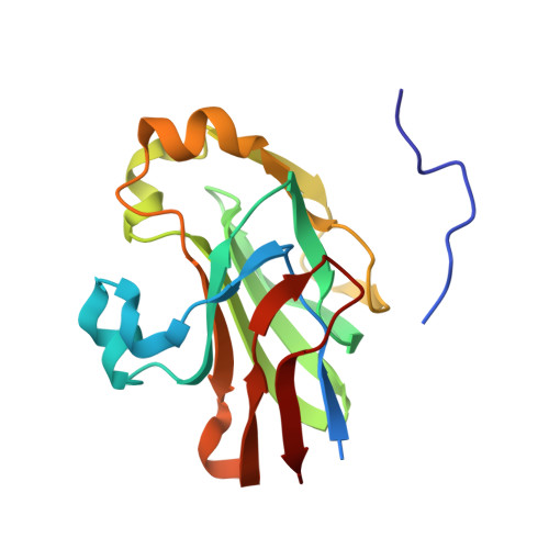
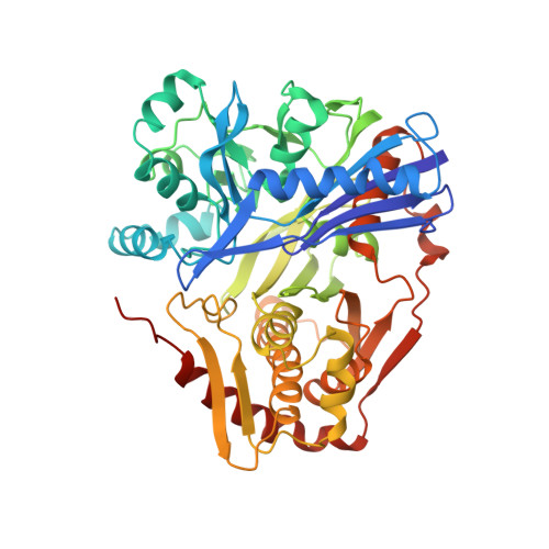
Structure of the regulatory complex of Escherichia coli IIIGlc with glycerol kinase.

Hurley, J.H.Faber, H.R.Worthylake, D.Meadow, N.D.Roseman, S.Pettigrew, D.W.Remington, S.J.

(1993) Science 259: 673-677

  • Primary Citation of Related Structures:  
    1GLA, 1GLB

  • PubMed Abstract: 

    The phosphocarrier protein IIIGlc is an integral component of the bacterial phosphotransferase (PTS) system. Unphosphorylated IIIGlc inhibits non-PTS carbohydrate transport systems by binding to diverse target proteins. The crystal structure at 2.6 A resolution of one of the targets, glycerol kinase (GK), in complex with unphosphorylated IIIGlc, glycerol, and adenosine diphosphate was determined. GK contains a region that is topologically identical to the adenosine triphosphate binding domains of hexokinase, the 70-kD heat shock cognate, and actin. IIIGlc binds far from the catalytic site of GK, indicating that long-range conformational changes mediate the inhibition of GK by IIIGlc. GK and IIIGlc are bound by hydrophobic and electrostatic interactions, with only one hydrogen bond involving an uncharged group. The phosphorylation site of IIIGlc, His90, is buried in a hydrophobic environment formed by the active site region of IIIGlc and a 3(10) helix of GK, suggesting that phosphorylation prevents IIIGlc binding to GK by directly disrupting protein-protein interactions.


  • Organizational Affiliation

    Institute of Molecular Biology, University of Oregon, Eugene 97403.


Macromolecules
Find similar proteins by:  (by identity cutoff)  |  3D Structure
Entity ID: 1
MoleculeChains Sequence LengthOrganismDetailsImage
GLUCOSE-SPECIFIC PROTEIN IIIGlcA [auth F]168Escherichia coliMutation(s): 0 
EC: 2.7.1.69
UniProt
Find proteins for P69783 (Escherichia coli (strain K12))
Explore P69783 
Go to UniProtKB:  P69783
Entity Groups  
Sequence Clusters30% Identity50% Identity70% Identity90% Identity95% Identity100% Identity
UniProt GroupP69783
Sequence Annotations
Expand
  • Reference Sequence
Find similar proteins by:  (by identity cutoff)  |  3D Structure
Entity ID: 2
MoleculeChains Sequence LengthOrganismDetailsImage
GLYCEROL KINASEB [auth G]501Escherichia coliMutation(s): 0 
EC: 2.7.1.30
UniProt
Find proteins for P0A6F3 (Escherichia coli (strain K12))
Explore P0A6F3 
Go to UniProtKB:  P0A6F3
Entity Groups  
Sequence Clusters30% Identity50% Identity70% Identity90% Identity95% Identity100% Identity
UniProt GroupP0A6F3
Sequence Annotations
Expand
  • Reference Sequence
Small Molecules
Ligands 2 Unique
IDChains Name / Formula / InChI Key2D Diagram3D Interactions
ADP
Query on ADP

Download Ideal Coordinates CCD File 
C [auth G]ADENOSINE-5'-DIPHOSPHATE
C10 H15 N5 O10 P2
XTWYTFMLZFPYCI-KQYNXXCUSA-N
GOL
Query on GOL

Download Ideal Coordinates CCD File 
D [auth G]GLYCEROL
C3 H8 O3
PEDCQBHIVMGVHV-UHFFFAOYSA-N
Experimental Data & Validation

Experimental Data

  • Method: X-RAY DIFFRACTION
  • Resolution: 2.60 Å
  • R-Value Work: 0.205 
  • R-Value Observed: 0.205 
  • Space Group: I 2 2 2
Unit Cell:
Length ( Å )Angle ( ˚ )
a = 123.7α = 90
b = 124.9β = 90
c = 134.2γ = 90
Software Package:
Software NamePurpose
X-PLORmodel building
X-PLORrefinement
X-PLORphasing

Structure Validation

View Full Validation Report


Entry History 

Deposition Data

Revision History  (Full details and data files)

  • Version 1.0: 1993-10-31
    Type: Initial release
  • Version 1.1: 2008-03-03
    Changes: Version format compliance
  • Version 1.2: 2011-07-13
    Changes: Non-polymer description, Version format compliance
  • Version 1.3: 2017-11-29
    Changes: Derived calculations, Other
  • Version 1.4: 2024-02-07
    Changes: Data collection, Database references, Derived calculations