Concanavalin a in a Dimeric Crystal Form: Revisiting Structural Accuracy and Molecular Flexibility
Kantardjieff, K., Hochtl, P., Segelke, B., Tao, F., Rupp, B.(2002) Acta Crystallogr D Biol Crystallogr 58: 735
- PubMed: 11976483
- DOI: https://doi.org/10.1107/s0907444901019588
- Primary Citation of Related Structures:
1GKB - PubMed Abstract:
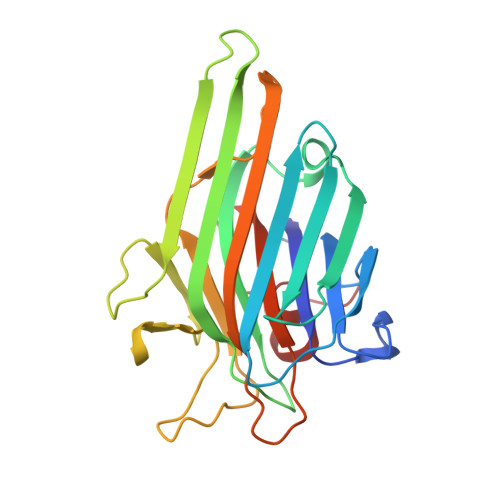
A structure of native concanavalin A (ConA), a hardy perennial of structural biology, has been determined in a dimeric crystal form at a resolution of 1.56 A (space group C222(1); unit-cell parameters a = 118.70, b = 101.38, c = 111.97 A; two molecules in the asymmetric unit). The structure has been refined to an R(free) of 0.206 (R = 0.178) after iterative model building and phase-bias removal using Shake&wARP. Correspondence between calculated water-tyrosine interactions and experimentally observed structures near the saccharide-binding site suggests that the observed interactions between Tyr12 and water in various crystal forms are to be expected and are not unique to the presence of an active site. The present structure differs from previously reported atomic resolution structures of ConA in several regions and extends insight into the conformational flexibility of this molecule. Furthermore, this third, low-temperature, structure of ConA in a different crystal form, independently refined using powerful model-bias removal techniques, affords the opportunity to revisit assessment of accuracy and precision in high- or atomic resolution protein structures. It is illustrated that several precise structures of the same molecule can differ substantially in local detail and users of crystallographic models are reminded to consider the potential impact when interpreting structures. Suggestions on how to effectively represent ensembles of crystallographic models of a given molecule are provided.
Organizational Affiliation:
Department of Chemistry and Biochemistry, W. M. Keck Foundation Center for Molecular Structure, California State University-Fullerton, Fullerton, CA 92834, USA.