1FWJ

KLEBSIELLA AEROGENES UREASE, NATIVE


Experimental Data Snapshot

  • Method: X-RAY DIFFRACTION
  • Resolution: 2.20 Å
  • R-Value Work: 0.173 
  • R-Value Observed: 0.173 

wwPDB Validation   3D Report Full Report


This is version 1.2 of the entry. See complete history


Literature

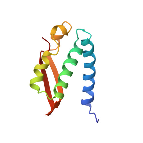
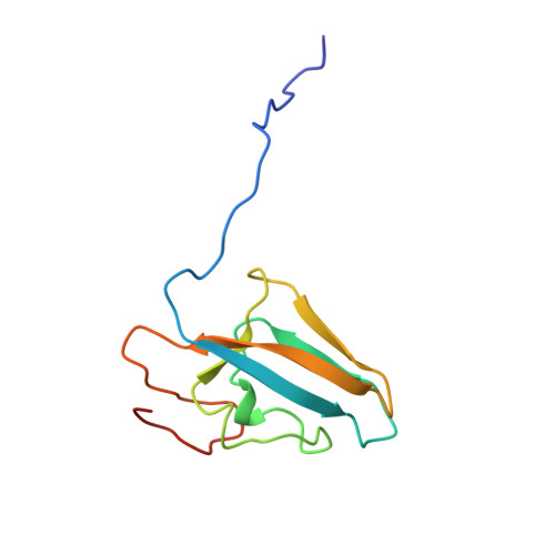
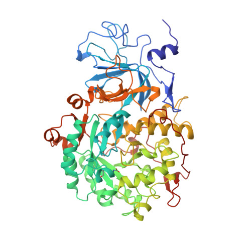
Structures of Cys319 variants and acetohydroxamate-inhibited Klebsiella aerogenes urease.

Pearson, M.A.Michel, L.O.Hausinger, R.P.Karplus, P.A.

(1997) Biochemistry 36: 8164-8172

  • DOI: https://doi.org/10.1021/bi970514j
  • Primary Citation of Related Structures:  
    1FWA, 1FWB, 1FWC, 1FWD, 1FWE, 1FWF, 1FWG, 1FWH, 1FWJ

  • PubMed Abstract: 

    Cys319 is located on a mobile flap covering the active site of Klebsiella aerogenes urease but does not play an essential role in catalysis. Four urease variants altered at position C319 range from having high activity (C319A) to no measurable activity (C319Y), indicating Cys is not required at this position, but its presence is highly influential [Martin, P. R., & Hausinger, R. P. (1992) J. Biol. Chem. 267, 20024-20027]. Here, we present 2.0 A resolution crystal structures of C319A, C319S, C319D, and C319Y proteins and the C319A variant inhibited by acetohydroxamic acid. These structures show changes in the hydration of the active site nickel ions and in the position and flexibility of the active site flap. The C319Y protein exhibits an alternate conformation of the flap, explaining its lack of activity. The changes in hydration and conformation suggest that there are suboptimal protein-solvent and protein-protein interactions in the empty urease active site which contribute to urease catalysis. Specifically, we hypothesize that the suboptimal interactions may provide a significant source of substrate binding energy, and such hidden energy may be a common phenomenon for enzymes that contain mobile active site loops and undergo an induced fit. The acetohydroxamic acid-bound structure reveals a chelate interaction similar to those seen in other metalloenzymes and in a small molecule nickel complex. The inhibitor binding mode supports the proposed mode of urea binding. We complement these structural studies with extended functional studies of C319A urease to show that it has enhanced stability and resistance to inhibition by buffers containing nickel ions. The near wild-type activity and enhanced stability of the C319A variant make it useful for further studies of urease structure-function relationships.


  • Organizational Affiliation

    Section of Biochemistry, Cornell University, Ithaca, New York 14853, USA.


Macromolecules
Find similar proteins by:  (by identity cutoff)  |  3D Structure
Entity ID: 1
MoleculeChains Sequence LengthOrganismDetailsImage
UREASE100Klebsiella aerogenesMutation(s): 0 
EC: 3.5.1.5
UniProt
Find proteins for P18316 (Klebsiella aerogenes)
Explore P18316 
Go to UniProtKB:  P18316
Entity Groups  
Sequence Clusters30% Identity50% Identity70% Identity90% Identity95% Identity100% Identity
UniProt GroupP18316
Sequence Annotations
Expand
  • Reference Sequence
Find similar proteins by:  (by identity cutoff)  |  3D Structure
Entity ID: 2
MoleculeChains Sequence LengthOrganismDetailsImage
UREASE106Klebsiella aerogenesMutation(s): 0 
EC: 3.5.1.5
UniProt
Find proteins for P18315 (Klebsiella aerogenes)
Explore P18315 
Go to UniProtKB:  P18315
Entity Groups  
Sequence Clusters30% Identity50% Identity70% Identity90% Identity95% Identity100% Identity
UniProt GroupP18315
Sequence Annotations
Expand
  • Reference Sequence
Find similar proteins by:  (by identity cutoff)  |  3D Structure
Entity ID: 3
MoleculeChains Sequence LengthOrganismDetailsImage
UREASE567Klebsiella aerogenesMutation(s): 1 
EC: 3.5.1.5
UniProt
Find proteins for P18314 (Klebsiella aerogenes)
Explore P18314 
Go to UniProtKB:  P18314
Entity Groups  
Sequence Clusters30% Identity50% Identity70% Identity90% Identity95% Identity100% Identity
UniProt GroupP18314
Sequence Annotations
Expand
  • Reference Sequence
Small Molecules
Ligands 1 Unique
IDChains Name / Formula / InChI Key2D Diagram3D Interactions
NI
Query on NI

Download Ideal Coordinates CCD File 
D [auth C],
E [auth C]
NICKEL (II) ION
Ni
VEQPNABPJHWNSG-UHFFFAOYSA-N
Modified Residues  1 Unique
IDChains TypeFormula2D DiagramParent
KCX
Query on KCX
C
L-PEPTIDE LINKINGC7 H14 N2 O4LYS
Experimental Data & Validation

Experimental Data

  • Method: X-RAY DIFFRACTION
  • Resolution: 2.20 Å
  • R-Value Work: 0.173 
  • R-Value Observed: 0.173 
  • Space Group: I 21 3
Unit Cell:
Length ( Å )Angle ( ˚ )
a = 170.8α = 90
b = 170.8β = 90
c = 170.8γ = 90
Software Package:
Software NamePurpose
X-PLORmodel building
X-PLORrefinement
X-PLORphasing

Structure Validation

View Full Validation Report


Entry History 

Deposition Data

Revision History  (Full details and data files)

  • Version 1.0: 1997-10-15
    Type: Initial release
  • Version 1.1: 2008-03-24
    Changes: Version format compliance
  • Version 1.2: 2011-07-13
    Changes: Derived calculations, Version format compliance