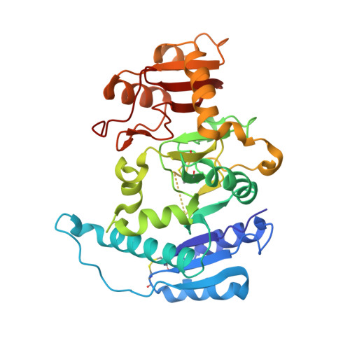
1FO8

CRYSTAL STRUCTURE OF N-ACETYLGLUCOSAMINYLTRANSFERASE I


Experimental Data Snapshot

  • Method: X-RAY DIFFRACTION
  • Resolution: 1.40 Å
  • R-Value Free: 0.185 
  • R-Value Work: 0.165 
  • R-Value Observed: 0.165 

wwPDB Validation   3D Report Full Report


This is version 1.2 of the entry. See complete history


Literature

X-ray crystal structure of rabbit N-acetylglucosaminyltransferase I: catalytic mechanism and a new protein superfamily.

Unligil, U.M.Zhou, S.Yuwaraj, S.Sarkar, M.Schachter, H.Rini, J.M.

(2000) EMBO J 19: 5269-5280

  • DOI: https://doi.org/10.1093/emboj/19.20.5269
  • Primary Citation of Related Structures:  
    1FO8, 1FO9, 1FOA

  • PubMed Abstract: 

    N:-acetylglucosaminyltransferase I (GnT I) serves as the gateway from oligomannose to hybrid and complex N:-glycans and plays a critical role in mammalian development and possibly all metazoans. We have determined the X-ray crystal structure of the catalytic fragment of GnT I in the absence and presence of bound UDP-GlcNAc/Mn(2+) at 1.5 and 1.8 A resolution, respectively. The structures identify residues critical for substrate binding and catalysis and provide evidence for similarity, at the mechanistic level, to the deglycosylation step of retaining beta-glycosidases. The structuring of a 13 residue loop, resulting from UDP-GlcNAc/Mn(2+) binding, provides an explanation for the ordered sequential 'Bi Bi' kinetics shown by GnT I. Analysis reveals a domain shared with Bacillus subtilis glycosyltransferase SpsA, bovine beta-1,4-galactosyl transferase 1 and Escherichia coli N:-acetylglucosamine-1-phosphate uridyltransferase. The low sequence identity, conserved fold and related functional features shown by this domain define a superfamily whose members probably share a common ancestor. Sequence analysis and protein threading show that the domain is represented in proteins from several glycosyltransferase families.


  • Organizational Affiliation

    Department of Medical Genetics and Microbiology, University of Toronto, Toronto, Ontario M5S 1A8, Canada.


Macromolecules
Find similar proteins by:  (by identity cutoff)  |  3D Structure
Entity ID: 1
MoleculeChains Sequence LengthOrganismDetailsImage
ALPHA-1,3-MANNOSYL-GLYCOPROTEIN BETA-1,2-N-ACETYLGLUCOSAMINYLTRANSFERASE343Oryctolagus cuniculusMutation(s): 0 
EC: 2.4.1.101
UniProt
Find proteins for P27115 (Oryctolagus cuniculus)
Explore P27115 
Go to UniProtKB:  P27115
Entity Groups  
Sequence Clusters30% Identity50% Identity70% Identity90% Identity95% Identity100% Identity
UniProt GroupP27115
Sequence Annotations
Expand
  • Reference Sequence
Small Molecules
Ligands 1 Unique
IDChains Name / Formula / InChI Key2D Diagram3D Interactions
MMC
Query on MMC

Download Ideal Coordinates CCD File 
B [auth A]METHYL MERCURY ION
C H3 Hg
DBUXSCUEGJMZAE-UHFFFAOYSA-N
Experimental Data & Validation

Experimental Data

  • Method: X-RAY DIFFRACTION
  • Resolution: 1.40 Å
  • R-Value Free: 0.185 
  • R-Value Work: 0.165 
  • R-Value Observed: 0.165 
  • Space Group: P 21 21 21
Unit Cell:
Length ( Å )Angle ( ˚ )
a = 40.382α = 90
b = 82.378β = 90
c = 102.487γ = 90
Software Package:
Software NamePurpose
DENZOdata reduction
SCALEPACKdata scaling
SOLVEphasing
CNSrefinement

Structure Validation

View Full Validation Report


Entry History 

Deposition Data

Revision History  (Full details and data files)

  • Version 1.0: 2001-04-25
    Type: Initial release
  • Version 1.1: 2008-04-27
    Changes: Version format compliance
  • Version 1.2: 2011-07-13
    Changes: Version format compliance