1FLM

DIMER OF FMN-BINDING PROTEIN FROM DESULFOVIBRIO VULGARIS (MIYAZAKI F)


Experimental Data Snapshot

  • Method: X-RAY DIFFRACTION
  • Resolution: 1.30 Å
  • R-Value Free: 0.182 
  • R-Value Observed: 0.150 

wwPDB Validation   3D Report Full Report


Ligand Structure Quality Assessment 


This is version 1.4 of the entry. See complete history


Literature

How do the x-ray structure and the NMR structure of FMN-binding protein differ?

Suto, K.Kawagoe, K.Shibata, N.Morimoto, Y.Higuchi, Y.Kitamura, M.Nakaya, T.Yasuoka, N.

(2000) Acta Crystallogr D Biol Crystallogr 56: 368-371

  • DOI: https://doi.org/10.1107/s0907444900000111
  • Primary Citation of Related Structures:  
    1FLM

  • PubMed Abstract: 

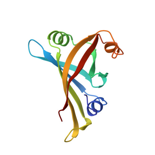
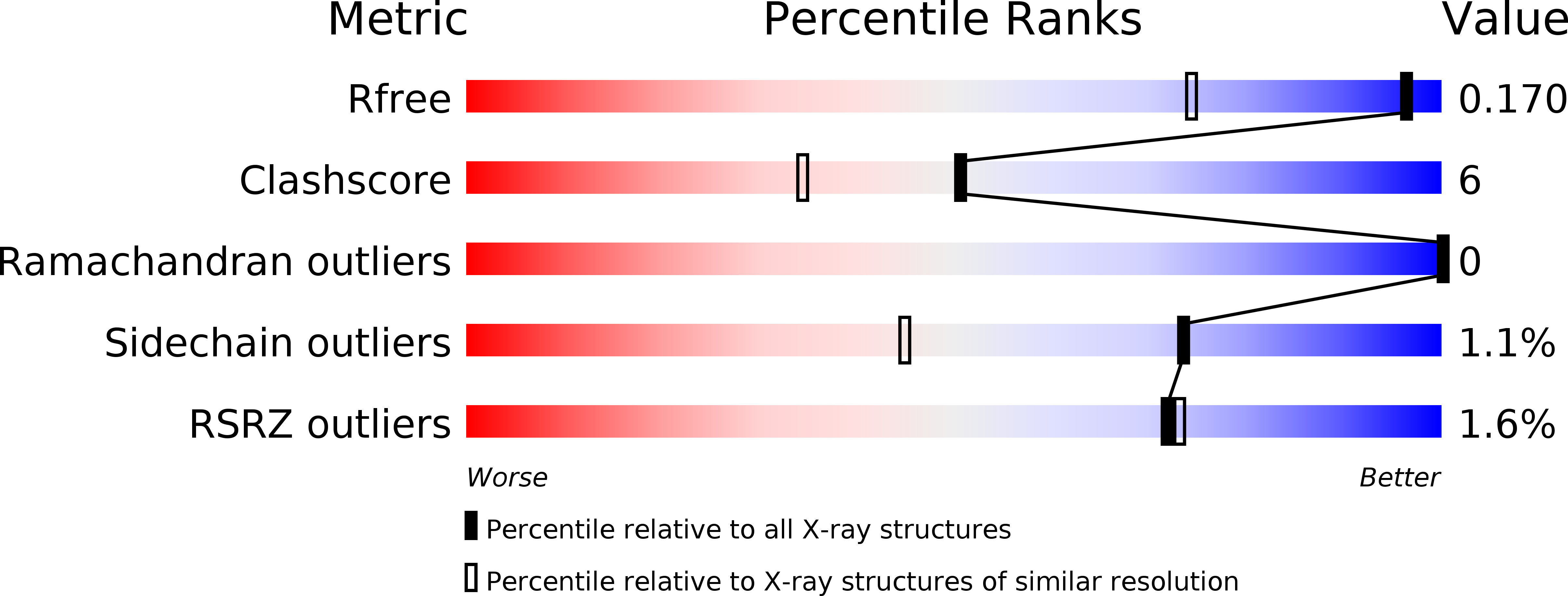
    The crystal structure of FMN-binding protein (FMN-bp) from Desulfovibrio vulgaris Miyazaki F was solved by the multiple isomorphous replacement method and refined to an R factor of 15.1% at 1.3 A resolution. FMN-bp exists in a dimeric form in the crystal, in contrast to the monomeric structure determined by NMR. R.m.s. deviations between the crystal structure and the solution structure are more than 2 A, which implies significant differences. There are some hydrophobic residues in the interface between the two monomers. In particular, Leu122 in the C-terminus has a close contact with the o-xylene moiety of FMN, while solvent molecules may cover the o-xylene moiety in the solution structure.


  • Organizational Affiliation

    Department of Life Science, Faculty of Science, Himeji Institute of Technology, 3-2-1 Kouto, Kamigori Ako-gun, Hyogo 678-1297, Japan.


Macromolecules
Find similar proteins by:  (by identity cutoff)  |  3D Structure
Entity ID: 1
MoleculeChains Sequence LengthOrganismDetailsImage
PROTEIN (FMN-BINDING PROTEIN)
A, B
122Nitratidesulfovibrio vulgaris str. 'Miyazaki FMutation(s): 0 
UniProt
Find proteins for Q46604 (Desulfovibrio vulgaris (strain DSM 19637 / Miyazaki F))
Explore Q46604 
Go to UniProtKB:  Q46604
Entity Groups  
Sequence Clusters30% Identity50% Identity70% Identity90% Identity95% Identity100% Identity
UniProt GroupQ46604
Sequence Annotations
Expand
  • Reference Sequence
Small Molecules
Binding Affinity Annotations 
IDSourceBinding Affinity
FMN PDBBind:  1FLM Kd: 0.43 (nM) from 1 assay(s)
Binding MOAD:  1FLM Kd: 0.43 (nM) from 1 assay(s)
Experimental Data & Validation

Experimental Data

  • Method: X-RAY DIFFRACTION
  • Resolution: 1.30 Å
  • R-Value Free: 0.182 
  • R-Value Observed: 0.150 
  • Space Group: P 1 21 1
Unit Cell:
Length ( Å )Angle ( ˚ )
a = 37.23α = 90
b = 84.6β = 94.1
c = 41.11γ = 90
Software Package:
Software NamePurpose
MLPHAREphasing
SHELXL-97refinement
DENZOdata reduction
SCALEPACKdata scaling

Structure Validation

View Full Validation Report


Ligand Structure Quality Assessment 


Entry History 

Deposition Data

Revision History  (Full details and data files)

  • Version 1.0: 2000-03-06
    Type: Initial release
  • Version 1.1: 2008-04-26
    Changes: Version format compliance
  • Version 1.2: 2011-07-13
    Changes: Version format compliance
  • Version 1.3: 2014-11-26
    Changes: Other
  • Version 1.4: 2023-12-27
    Changes: Data collection, Database references, Derived calculations