X-ray structure of the quinoprotein ethanol dehydrogenase from Pseudomonas aeruginosa: basis of substrate specificity.
Keitel, T., Diehl, A., Knaute, T., Stezowski, J.J., Hohne, W., Gorisch, H.(2000) J Mol Biol 297: 961-974
- PubMed: 10736230
- DOI: https://doi.org/10.1006/jmbi.2000.3603
- Primary Citation of Related Structures:
1FLG - PubMed Abstract:
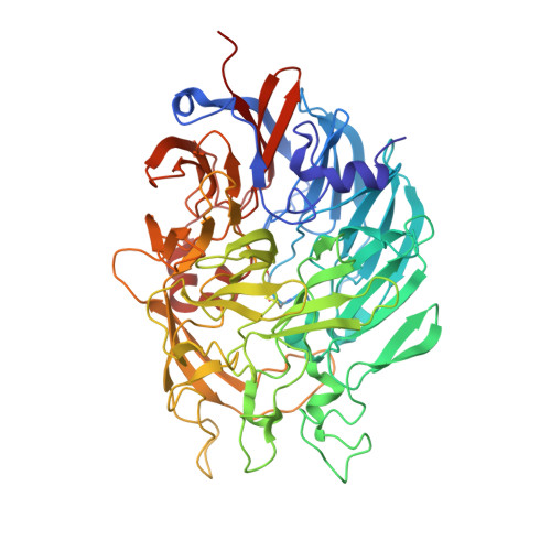
The homodimeric enzyme form of quinoprotein ethanol dehydrogenase from Pseudomonas aeruginosa ATCC 17933 crystallizes readily with the space group R3. The X-ray structure was solved at 2.6 A resolution by molecular replacement. Aside from differences in some loops, the folding of the enzyme is very similar to the large subunit of the quinoprotein methanol dehydrogenases from Methylobacterium extorquens or Methylophilus W3A1. Eight W-shaped beta-sheet motifs are arranged circularly in a propeller-like fashion forming a disk-shaped superbarrel. No electron density for a small subunit like that in methanol dehydrogenase could be found. The prosthetic group is located in the centre of the superbarrel and is coordinated to a calcium ion. Most amino acid residues found in close contact with the prosthetic group pyrroloquinoline quinone and the Ca(2+) are conserved between the quinoprotein ethanol dehydrogenase structure and that of the methanol dehydrogenases. The main differences in the active-site region are a bulky tryptophan residue in the active-site cavity of methanol dehydrogenase, which is replaced by a phenylalanine and a leucine side-chain in the ethanol dehydrogenase structure and a leucine residue right above the pyrrolquinoline quinone group in methanol dehydrogenase which is replaced by a tryptophan side-chain. Both amino acid exchanges appear to have an important influence, causing different substrate specificities of these otherwise very similar enzymes. In addition to the Ca(2+) in the active-site cavity found also in methanol dehydrogenase, ethanol dehydrogenase contains a second Ca(2+)-binding site at the N terminus, which contributes to the stability of the native enzyme.
Organizational Affiliation:
Universitätsklinikum Charité Institut für Biochemie, Humboldt-Universität zu Berlin, Monbijoustr. 2, Berlin, D-10117, Germany.