1FIG

ROUTES TO CATALYSIS: STRUCTURE OF A CATALYTIC ANTIBODY AND COMPARISON WITH ITS NATURAL COUNTERPART


Experimental Data Snapshot

  • Method: X-RAY DIFFRACTION
  • Resolution: 3.00 Å
  • R-Value Work: 0.220 
  • R-Value Observed: 0.220 

wwPDB Validation   3D Report Full Report


This is version 1.2 of the entry. See complete history


Literature

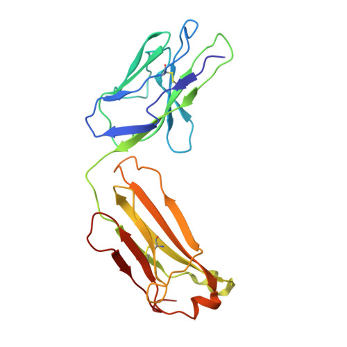
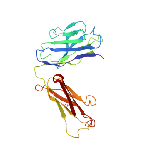
Routes to catalysis: structure of a catalytic antibody and comparison with its natural counterpart.

Haynes, M.R.Stura, E.A.Hilvert, D.Wilson, I.A.

(1994) Science 263: 646-652

  • DOI: https://doi.org/10.1126/science.8303271
  • Primary Citation of Related Structures:  
    1FIG

  • PubMed Abstract: 

    The three-dimensional structure of a catalytic antibody (1F7) with chorismate mutase activity has been determined to 3.0 A resolution as a complex with a transition state analog. The structural data suggest that the antibody stabilizes the same conformationally restricted pericyclic transition state as occurs in the uncatalyzed reaction. Overall shape and charge complementarity between the combining site and the transition state analog dictate preferential binding of the correct substrate enantiomer in a conformation appropriate for reaction. Comparison with the structure of a chorismate mutase enzyme indicates an overall similarity between the catalytic mechanism employed by the two proteins. Differences in the number of specific interactions available for restricting the rotational degrees of freedom in the transition state, and the lack of multiple electrostatic interactions that might stabilize charge separation in this highly polarized metastable species, are likely to account for the observed 10(4) times lower activity of the antibody relative to that of the natural enzymes that catalyze this reaction. The structure of the 1F7 Fab'-hapten complex provides confirmation that the properties of an antibody catalyst faithfully reflect the design of the transition state analog.


  • Organizational Affiliation

    Department of Molecular Biology, Scripps Research Institute, La Jolla, CA 92037.


Macromolecules
Find similar proteins by:  (by identity cutoff)  |  3D Structure
Entity ID: 1
MoleculeChains Sequence LengthOrganismDetailsImage
IGG1-KAPPA 1F7 FAB (LIGHT CHAIN)A [auth L]215Mus musculusMutation(s): 0 
Entity Groups  
Sequence Clusters30% Identity50% Identity70% Identity90% Identity95% Identity100% Identity
Sequence Annotations
Expand
  • Reference Sequence
Find similar proteins by:  (by identity cutoff)  |  3D Structure
Entity ID: 2
MoleculeChains Sequence LengthOrganismDetailsImage
IGG1-KAPPA 1F7 FAB (HEAVY CHAIN)B [auth H]216Mus musculusMutation(s): 0 
Entity Groups  
Sequence Clusters30% Identity50% Identity70% Identity90% Identity95% Identity100% Identity
Sequence Annotations
Expand
  • Reference Sequence
Small Molecules
Ligands 1 Unique
IDChains Name / Formula / InChI Key2D Diagram3D Interactions
TSA
Query on TSA

Download Ideal Coordinates CCD File 
C [auth H]8-HYDROXY-2-OXA-BICYCLO[3.3.1]NON-6-ENE-3,5-DICARBOXYLIC ACID
C10 H12 O6
KRZHNRULRHECRF-JQCUSGDOSA-N
Binding Affinity Annotations 
IDSourceBinding Affinity
TSA PDBBind:  1FIG Ki: 600 (nM) from 1 assay(s)
Experimental Data & Validation

Experimental Data

  • Method: X-RAY DIFFRACTION
  • Resolution: 3.00 Å
  • R-Value Work: 0.220 
  • R-Value Observed: 0.220 
  • Space Group: P 21 21 21
Unit Cell:
Length ( Å )Angle ( ˚ )
a = 37.1α = 90
b = 63.3β = 90
c = 178.5γ = 90
Software Package:
Software NamePurpose
X-PLORmodel building
X-PLORrefinement
X-PLORphasing

Structure Validation

View Full Validation Report


Entry History 

Deposition Data

Revision History  (Full details and data files)

  • Version 1.0: 1994-05-31
    Type: Initial release
  • Version 1.1: 2008-03-03
    Changes: Version format compliance
  • Version 1.2: 2011-07-13
    Changes: Version format compliance