1FE8

CRYSTAL STRUCTURE OF THE VON WILLEBRAND FACTOR A3 DOMAIN IN COMPLEX WITH A FAB FRAGMENT OF IGG RU5 THAT INHIBITS COLLAGEN BINDING


Experimental Data Snapshot

  • Method: X-RAY DIFFRACTION
  • Resolution: 2.03 Å
  • R-Value Free: 0.264 
  • R-Value Work: 0.227 
  • R-Value Observed: 0.229 

wwPDB Validation   3D Report Full Report


Ligand Structure Quality Assessment 


This is version 2.0 of the entry. See complete history


Literature

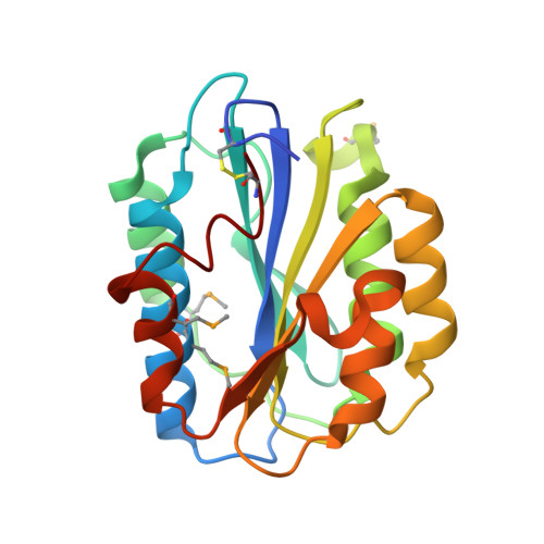
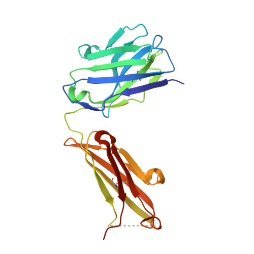
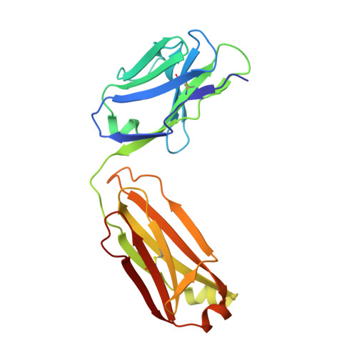
Identification of the collagen-binding site of the von Willebrand factor A3-domain.

Romijn, R.A.Bouma, B.Wuyster, W.Gros, P.Kroon, J.Sixma, J.J.Huizinga, E.G.

(2001) J Biol Chem 276: 9985-9991

  • DOI: https://doi.org/10.1074/jbc.M006548200
  • Primary Citation of Related Structures:  
    1FE8

  • PubMed Abstract: 

    Von Willebrand factor (vWF) is a multimeric glycoprotein that mediates platelet adhesion and thrombus formation at sites of vascular injury. vWF functions as a molecular bridge between collagen and platelet receptor glycoprotein Ib. The major collagen-binding site of vWF is contained within the A3 domain, but its precise location is unknown. To localize the collagen-binding site, we determined the crystal structure of A3 in complex with an Fab fragment of antibody RU5 that inhibits collagen binding. The structure shows that RU5 recognizes a nonlinear epitope consisting of residues 962-966, 981-997, and 1022-1026. Alanine mutants were constructed of residues Arg(963), Glu(987), His(990), Arg(1016), and His(1023), located in or close to the epitope. Mutants were expressed as fully processed multimeric vWF. Mutation of His(1023) abolished collagen binding, whereas mutation of Arg(963) and Arg(1016) reduced collagen binding by 25-35%. These residues are part of loops alpha3beta4 and alpha1beta2 and alpha-helix 3, respectively, and lie near the bottom face of the domain. His(1023) and flanking residues display multiple conformations in available A3-crystal structures, suggesting that binding of A3 to collagen involves an induced-fit mechanism. The collagen-binding site of A3 is located distant from the top face of the domain where collagen-binding sites are found in homologous integrin I domains.


  • Organizational Affiliation

    Thrombosis and Haemostasis Laboratory, Department of Haematology, University Medical Center and Institute of Biomembranes, HP G03.647, P. O. Box 85500, 3508 GA Utrecht, The Netherlands.


Macromolecules
Find similar proteins by:  (by identity cutoff)  |  3D Structure
Entity ID: 1
MoleculeChains Sequence LengthOrganismDetailsImage
VON WILLEBRAND FACTORA,
D [auth B],
G [auth C]
196Homo sapiensMutation(s): 4 
UniProt & NIH Common Fund Data Resources
Find proteins for P04275 (Homo sapiens)
Explore P04275 
Go to UniProtKB:  P04275
PHAROS:  P04275
GTEx:  ENSG00000110799 
Entity Groups  
Sequence Clusters30% Identity50% Identity70% Identity90% Identity95% Identity100% Identity
UniProt GroupP04275
Sequence Annotations
Expand
  • Reference Sequence
Find similar proteins by:  (by identity cutoff)  |  3D Structure
Entity ID: 2
MoleculeChains Sequence LengthOrganismDetailsImage
IMMUNOGLOBULIN IGG RU5B [auth H],
E [auth I],
H [auth J]
210Mus musculusMutation(s): 0 
UniProt
Find proteins for P01863 (Mus musculus)
Explore P01863 
Go to UniProtKB:  P01863
Entity Groups  
Sequence Clusters30% Identity50% Identity70% Identity90% Identity95% Identity100% Identity
UniProt GroupP01863
Sequence Annotations
Expand
  • Reference Sequence
Find similar proteins by:  (by identity cutoff)  |  3D Structure
Entity ID: 3
MoleculeChains Sequence LengthOrganismDetailsImage
IMMUNOGLOBULIN IGG RU5C [auth L],
F [auth M],
I [auth N]
211Mus musculusMutation(s): 0 
Entity Groups  
Sequence Clusters30% Identity50% Identity70% Identity90% Identity95% Identity100% Identity
Sequence Annotations
Expand
  • Reference Sequence
Oligosaccharides

Help

Entity ID: 4
MoleculeChains Length2D Diagram Glycosylation3D Interactions
alpha-L-fucopyranose-(1-6)-2-acetamido-2-deoxy-beta-D-glucopyranoseJ [auth D],
K [auth E],
L [auth F]
2N-Glycosylation
Glycosylation Resources
GlyTouCan:  G86851RC
GlyCosmos:  G86851RC
GlyGen:  G86851RC
Small Molecules
Modified Residues  1 Unique
IDChains TypeFormula2D DiagramParent
MSE
Query on MSE
A,
D [auth B],
G [auth C]
L-PEPTIDE LINKINGC5 H11 N O2 SeMET
Binding Affinity Annotations 
IDSourceBinding Affinity
RU5 PDBBind:  1FE8 Kd: 2000 (nM) from 1 assay(s)
Experimental Data & Validation

Experimental Data

  • Method: X-RAY DIFFRACTION
  • Resolution: 2.03 Å
  • R-Value Free: 0.264 
  • R-Value Work: 0.227 
  • R-Value Observed: 0.229 
  • Space Group: C 1 2 1
Unit Cell:
Length ( Å )Angle ( ˚ )
a = 121.8α = 90
b = 183.6β = 116.2
c = 131.8γ = 90
Software Package:
Software NamePurpose
DENZOdata reduction
SCALEPACKdata scaling
AMoREphasing
CNSrefinement
CNSphasing

Structure Validation

View Full Validation Report


Ligand Structure Quality Assessment 


Entry History 

Deposition Data

Revision History  (Full details and data files)

  • Version 1.0: 2001-04-04
    Type: Initial release
  • Version 1.1: 2008-04-27
    Changes: Version format compliance
  • Version 1.2: 2011-07-13
    Changes: Non-polymer description, Version format compliance
  • Version 2.0: 2020-07-29
    Type: Remediation
    Reason: Carbohydrate remediation
    Changes: Advisory, Atomic model, Data collection, Database references, Derived calculations, Structure summary