Solution structure of the Cu(I) and apo forms of the yeast metallochaperone, Atx1.
Arnesano, F., Banci, L., Bertini, I., Huffman, D.L., O'Halloran, T.V.(2001) Biochemistry 40: 1528-1539
- PubMed: 11327811
- DOI: https://doi.org/10.1021/bi0014711
- Primary Citation of Related Structures:
1FD8, 1FES - PubMed Abstract:
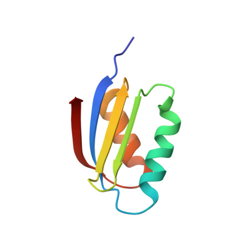
The (1)H NMR solution structure of the Cu(I)-bound form of Atx1, a 73-amino acid metallochaperone protein from the yeast Saccharomyces cerevisiae, has been determined. Ninety percent of the (1)H and 95% of the (15)N resonances were assigned, and 1184 meaningful NOEs and 42 (3)J(HNH)(alpha) and 60 (1)J(HN) residual dipolar couplings provided a family of structures with rmsd values to the mean structure of 0.37 +/- 0.07 A for the backbone and 0.83 +/- 0.08 A for all heavy atoms. The structure is constituted by four antiparallel beta strands and two alpha helices in a betaalphabetabetaalphabeta fold. Following EXAFS data [Pufahl, R., Singer, C. P., Peariso, K. L., Lin, S.-J., Schmidt, P. J., Fahrni, C. J., Cizewski Culotta, V., Penner-Hahn, J. E., and O'Halloran, T. V. (1997) Science 278, 853-856], a copper ion can be placed between two sulfur atoms of Cys15 and Cys18. The structure of the reduced apo form has also been determined with similar resolution using 1252 meaningful NOEs (rmsd values for the family to the mean structure are 0.67 +/- 0.12 A for the backbone and 1.00 +/- 0.12 A for all heavy atoms). Comparison of the Cu(I) and apo conformations of the protein reveals that the Cu(I) binding cysteines move from a buried site in the bound metal form to a solvent-exposed conformation on the surface of the protein after copper release. Furthermore, copper release leads to a less helical character in the metal binding site. Comparison with the Hg(II)-Atx1 solid-state structure [Rosenzweig, A. C., Huffman, D. L., Hou, M. Y., Wernimont, A. K., Pufahl, R. A., and O'Halloran, T. V. (1999) Structure 7, 605-617] provides insights into the copper transfer mechanism, and a pivotal role for Lys65 in the metal capture and release process is proposed.
Organizational Affiliation:
Magnetic Resonance Center CERM, University of Florence, Via Luigi Sacconi 6, 50019, Sesto Fiorentino, Florence, Italy.