


Structure Comparison of Two Platelet Factor 4 Mutants with the Wild-type Reveals the Epitopes for the Heparin-induced Thrombocytopenia Antibodies
Yang, J., Doyle, M., Faulk, T., Visentin, G., Aster, R., Edwards, B.To be published.
Experimental Data Snapshot
wwPDB Validation 3D Report Full Report

Entity ID: 1 | |||||
|---|---|---|---|---|---|
| Molecule | Chains | Sequence Length | Organism | Details | Image |
| PLATELET FACTOR 4 | 70 | Homo sapiens | Mutation(s): 3 | 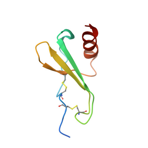 | |
UniProt & NIH Common Fund Data Resources | |||||
Find proteins for P02776 (Homo sapiens) Explore P02776 Go to UniProtKB: P02776 | |||||
PHAROS: P02776 GTEx: ENSG00000163737 | |||||
Entity Groups | |||||
| Sequence Clusters | 30% Identity50% Identity70% Identity90% Identity95% Identity100% Identity | ||||
| UniProt Group | P02776 | ||||
Sequence AnnotationsExpand | |||||
| |||||
| Length ( Å ) | Angle ( ˚ ) |
|---|---|
| a = 81.82 | α = 90 |
| b = 77.48 | β = 90 |
| c = 43.25 | γ = 90 |
| Software Name | Purpose |
|---|---|
| SCALEPACK | data scaling |
| CNS | refinement |
| CNS | phasing |
RCSB PDB (citation) is hosted by
RCSB PDB is a member of the

