1F1E

CRYSTAL STRUCTURE OF THE HISTONE FROM METHANOPYRUS KANDLERI


Experimental Data Snapshot

  • Method: X-RAY DIFFRACTION
  • Resolution: 1.37 Å
  • R-Value Free: 0.206 
  • R-Value Work: 0.159 
  • R-Value Observed: 0.159 

wwPDB Validation   3D Report Full Report


This is version 1.3 of the entry. See complete history


Literature

An ancestral nuclear protein assembly: crystal structure of the Methanopyrus kandleri histone.

Fahrner, R.L.Cascio, D.Lake, J.A.Slesarev, A.

(2001) Protein Sci 10: 2002-2007

  • DOI: https://doi.org/10.1110/ps.10901
  • Primary Citation of Related Structures:  
    1F1E

  • PubMed Abstract: 

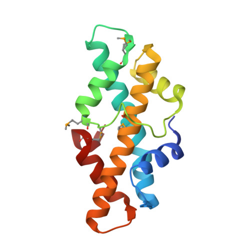
    Eukaryotic histone proteins condense DNA into compact structures called nucleosomes. Nucleosomes were viewed as a distinguishing feature of eukaryotes prior to identification of histone orthologs in methanogens. Although evolutionarily distinct from methanogens, the methane-producing hyperthermophile Methanopyrus kandleri produces a novel, 154-residue histone (HMk). Amino acid sequence comparisons show that HMk differs from both methanogenic and eukaryotic histones, in that it contains two histone-fold ms within a single chain. The two HMk histone-fold ms, N and C terminal, are 28% identical in amino acid sequence to each other and approximately 21% identical in amino acid sequence to other histone proteins. Here we present the 1.37-A-resolution crystal structure of HMk and report that the HMk monomer structure is homologous to the eukaryotic histone heterodimers. In the crystal, HMk forms a dimer homologous to [H3-H4](2) in the eukaryotic nucleosome. Based on the spatial similarities to structural ms found in the eukaryotic nucleosome that are important for DNA-binding, we infer that the Methanopyrus histone binds DNA in a manner similar to the eukaryotic histone tetramer [H3-H4](2).


  • Organizational Affiliation

    Department of Chemistry and Biochemistry, University of California, Los Angeles, California 90095-1570, USA.


Macromolecules
Find similar proteins by:  (by identity cutoff)  |  3D Structure
Entity ID: 1
MoleculeChains Sequence LengthOrganismDetailsImage
HISTONE FOLD PROTEIN154Methanopyrus kandleriMutation(s): 4 
UniProt
Find proteins for O93641 (Methanopyrus kandleri)
Explore O93641 
Go to UniProtKB:  O93641
Entity Groups  
Sequence Clusters30% Identity50% Identity70% Identity90% Identity95% Identity100% Identity
UniProt GroupO93641
Sequence Annotations
Expand
  • Reference Sequence
Small Molecules
Ligands 1 Unique
IDChains Name / Formula / InChI Key2D Diagram3D Interactions
CL
Query on CL

Download Ideal Coordinates CCD File 
B [auth A]CHLORIDE ION
Cl
VEXZGXHMUGYJMC-UHFFFAOYSA-M
Modified Residues  1 Unique
IDChains TypeFormula2D DiagramParent
MSE
Query on MSE
A
L-PEPTIDE LINKINGC5 H11 N O2 SeMET
Experimental Data & Validation

Experimental Data

  • Method: X-RAY DIFFRACTION
  • Resolution: 1.37 Å
  • R-Value Free: 0.206 
  • R-Value Work: 0.159 
  • R-Value Observed: 0.159 
  • Space Group: P 41 21 2
Unit Cell:
Length ( Å )Angle ( ˚ )
a = 57.4α = 90
b = 57.4β = 90
c = 96.95γ = 90
Software Package:
Software NamePurpose
SCALEPACKdata scaling
SOLVEphasing
SHELXL-97refinement

Structure Validation

View Full Validation Report


Entry History 

Deposition Data

Revision History  (Full details and data files)

  • Version 1.0: 2001-10-31
    Type: Initial release
  • Version 1.1: 2008-04-27
    Changes: Version format compliance
  • Version 1.2: 2011-07-13
    Changes: Derived calculations, Version format compliance
  • Version 1.3: 2017-10-04
    Changes: Refinement description