1EEY

Crystal Structure Determination Of HLA A2 Complexed to Peptide GP2 with the substitution (I2L/V5L/L9V)


Experimental Data Snapshot

  • Method: X-RAY DIFFRACTION
  • Resolution: 2.25 Å
  • R-Value Free: 0.286 
  • R-Value Work: 0.248 
  • R-Value Observed: 0.248 

wwPDB Validation   3D Report Full Report


This is version 1.3 of the entry. See complete history


Literature

Class I major histocompatibility complex anchor substitutions alter the conformation of T cell receptor contacts.

Sharma, A.K.Kuhns, J.J.Yan, S.Friedline, R.H.Long, B.Tisch, R.Collins, E.J.

(2001) J Biol Chem 276: 21443-21449

  • DOI: https://doi.org/10.1074/jbc.M010791200
  • Primary Citation of Related Structures:  
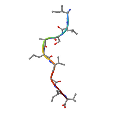
    1EEY, 1EEZ

  • PubMed Abstract: 

    An immunogenic peptide (GP2) derived from HER-2/neu binds to HLA-A2.1 very poorly. Some altered-peptide ligands (APL) of GP2 have increased binding affinity and generate improved cytotoxic T lymphocyte recognition of GP2-presenting tumor cells, but most do not. Increases in binding affinity of single-substitution APL are not additive in double-substitution APL. A common first assumption about peptide binding to class I major histocompatibility complex is that each residue binds independently. In addition, immunologists interested in immunotherapy frequently assume that anchor substitutions do not affect T cell receptor contact residues. However, the crystal structures of two GP2 APL show that the central residues change position depending on the identity of the anchor residue(s). Thus, it is clear that subtle changes in the identity of anchor residues may have significant effects on the positions of the T cell receptor contact residues.


  • Organizational Affiliation

    Departments of Microbiology, the Lineberger Comprehensive Cancer Center, University of North Carolina, Chapel Hill, North Carolina 27599, USA.


Macromolecules
Find similar proteins by:  (by identity cutoff)  |  3D Structure
Entity ID: 1
MoleculeChains Sequence LengthOrganismDetailsImage
HLA-A2.1 MHC CLASS I (HEAVY CHAIN)
A, D
275Homo sapiensMutation(s): 0 
UniProt & NIH Common Fund Data Resources
Find proteins for P04439 (Homo sapiens)
Explore P04439 
Go to UniProtKB:  P04439
PHAROS:  P04439
GTEx:  ENSG00000206503 
Entity Groups  
Sequence Clusters30% Identity50% Identity70% Identity90% Identity95% Identity100% Identity
UniProt GroupP04439
Sequence Annotations
Expand
  • Reference Sequence
Find similar proteins by:  (by identity cutoff)  |  3D Structure
Entity ID: 2
MoleculeChains Sequence LengthOrganismDetailsImage
BETA-2-MICROGLOBULIN (LIGHT CHAIN)
B, E
100Homo sapiensMutation(s): 0 
UniProt & NIH Common Fund Data Resources
Find proteins for P61769 (Homo sapiens)
Explore P61769 
Go to UniProtKB:  P61769
PHAROS:  P61769
GTEx:  ENSG00000166710 
Entity Groups  
Sequence Clusters30% Identity50% Identity70% Identity90% Identity95% Identity100% Identity
UniProt GroupP61769
Sequence Annotations
Expand
  • Reference Sequence

Find similar proteins by:  Sequence   |   3D Structure  

Entity ID: 3
MoleculeChains Sequence LengthOrganismDetailsImage
GP2 PEPTIDE
C, F
9N/AMutation(s): 0 
Sequence Annotations
Expand
  • Reference Sequence
Experimental Data & Validation

Experimental Data

  • Method: X-RAY DIFFRACTION
  • Resolution: 2.25 Å
  • R-Value Free: 0.286 
  • R-Value Work: 0.248 
  • R-Value Observed: 0.248 
  • Space Group: P 1
Unit Cell:
Length ( Å )Angle ( ˚ )
a = 49.95α = 82.07
b = 62.93β = 76.5
c = 74.65γ = 78.04
Software Package:
Software NamePurpose
DENZOdata reduction
SCALEPACKdata scaling
AMoREphasing
CNSrefinement

Structure Validation

View Full Validation Report


Entry History 

Deposition Data

Revision History  (Full details and data files)

  • Version 1.0: 2003-06-10
    Type: Initial release
  • Version 1.1: 2008-04-27
    Changes: Version format compliance
  • Version 1.2: 2011-07-13
    Changes: Version format compliance
  • Version 1.3: 2022-12-21
    Changes: Database references