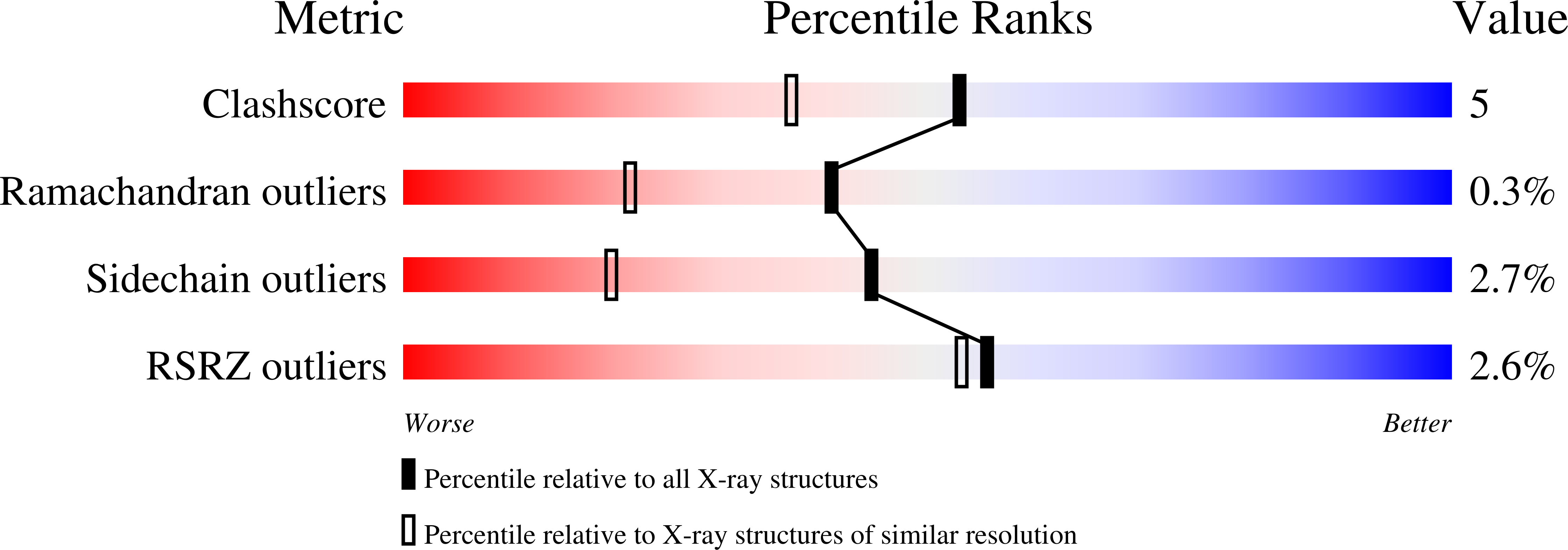
Crystal structure of the catalytic domain of a bacterial cellulase belonging to family 5.
Ducros, V., Czjzek, M., Belaich, A., Gaudin, C., Fierobe, H.P., Belaich, J.P., Davies, G.J., Haser, R.(1995) Structure 3: 939-949
- PubMed: 8535787
- DOI: https://doi.org/10.1016/S0969-2126(01)00228-3
- Primary Citation of Related Structures:
1EDG - PubMed Abstract:
Cellulases are glycosyl hydrolases--enzymes that hydrolyze glycosidic bonds. They have been widely studied using biochemical and microbiological techniques and have attracted industrial interest because of their potential in biomass conversion and in the paper and textile industries. Glycosyl hydrolases have lately been assigned to specific families on the basis of similarities in their amino acid sequences. The cellulase endoglucanase A produced by Clostridium cellulolyticum (CelCCA) belongs to family 5. We have determined the crystal structure of the catalytic domain of CelCCA at a resolution of 2.4 A and refined it to 1.6 A. The structure was solved by the multiple isomorphous replacement method. The overall structural fold, (alpha/beta)8, belongs to the TIM barrel motif superfamily. The catalytic centre is located at the C-terminal ends of the beta strands; the aromatic residues, forming the substrate-binding site, are arranged along a long cleft on the surface of the globular enzyme. Strictly conserved residues within family 5 are described with respect to their catalytic function. The proton donor, Glu170, and the nucleophile, Glu307, are localized on beta strands IV and VII, respectively, and are separated by 5.5 A, as expected for enzymes which retain the configuration of the substrate's anomeric carbon. Structure determination of the catalytic domain of CelCCA allows a comparison with related enzymes belonging to glycosyl hydrolase families 2, 10 and 17, which also display an (alpha/beta)8 fold.
Organizational Affiliation:
Institut de Biologie Structurale et Microbiologie, URA 1296, CNRS, Marseille, France.


