1DKR

CRYSTAL STRUCTURES OF BACILLUS SUBTILIS PHOSPHORIBOSYLPYROPHOSPHATE SYNTHETASE: MOLECULAR BASIS OF ALLOSTERIC INHIBITION AND ACTIVATION.


Experimental Data Snapshot

  • Method: X-RAY DIFFRACTION
  • Resolution: 2.30 Å
  • R-Value Free: 0.240 
  • R-Value Work: 0.180 
  • R-Value Observed: 0.180 

wwPDB Validation   3D Report Full Report


This is version 1.4 of the entry. See complete history


Literature

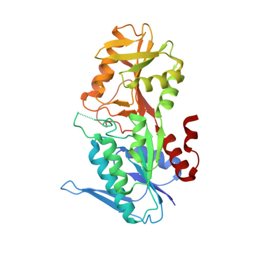
Structural basis for the function of Bacillus subtilis phosphoribosyl-pyrophosphate synthetase.

Eriksen, T.A.Kadziola, A.Bentsen, A.K.Harlow, K.W.Larsen, S.

(2000) Nat Struct Biol 7: 303-308

  • DOI: https://doi.org/10.1038/74069
  • Primary Citation of Related Structures:  
    1DKR, 1DKU

  • PubMed Abstract: 

    Here we report the first three-dimensional structure of a phosphoribosylpyrophosphate (PRPP) synthetase. PRPP is an essential intermediate in several biosynthetic pathways. Structures of the Bacillus subtilis PRPP synthetase in complex with analogs of the activator phosphate and the allosteric inhibitor ADP show that the functional form of the enzyme is a hexamer. The individual subunits fold into two domains, both of which resemble the type I phosphoribosyltransfereases. The active site is located between the two domains and includes residues from two subunits. Phosphate and ADP bind to the same regulatory site consisting of residues from three subunits of the hexamer. In addition to identifying residues important for binding substrates and effectors, the structures suggest a novel mode of allosteric regulation.


  • Organizational Affiliation

    Center for Crystallographic Studies, Department of Chemistry, University of Copenhagen, Universitetsparken 5, DK-2100 Copenhagen, Denmark.


Macromolecules
Find similar proteins by:  (by identity cutoff)  |  3D Structure
Entity ID: 1
MoleculeChains Sequence LengthOrganismDetailsImage
PHOSPHORIBOSYL PYROPHOSPHATE SYNTHETASE
A, B
317Bacillus subtilisMutation(s): 0 
EC: 2.7.6.1
UniProt
Find proteins for P14193 (Bacillus subtilis (strain 168))
Explore P14193 
Go to UniProtKB:  P14193
Entity Groups  
Sequence Clusters30% Identity50% Identity70% Identity90% Identity95% Identity100% Identity
UniProt GroupP14193
Sequence Annotations
Expand
  • Reference Sequence
Experimental Data & Validation

Experimental Data

  • Method: X-RAY DIFFRACTION
  • Resolution: 2.30 Å
  • R-Value Free: 0.240 
  • R-Value Work: 0.180 
  • R-Value Observed: 0.180 
  • Space Group: P 63
Unit Cell:
Length ( Å )Angle ( ˚ )
a = 115.61α = 90
b = 115.61β = 90
c = 107.75γ = 120
Software Package:
Software NamePurpose
MLPHAREphasing
DMmodel building
X-PLORrefinement
DENZOdata reduction
CCP4data scaling
ROTAVATAdata scaling
TRUNCATEdata scaling
DMphasing

Structure Validation

View Full Validation Report


Entry History 

Deposition Data

Revision History  (Full details and data files)

  • Version 1.0: 2000-04-05
    Type: Initial release
  • Version 1.1: 2008-04-27
    Changes: Version format compliance
  • Version 1.2: 2011-07-13
    Changes: Derived calculations, Version format compliance
  • Version 1.3: 2017-10-04
    Changes: Refinement description
  • Version 1.4: 2024-02-07
    Changes: Data collection, Database references, Derived calculations