1DI9

THE STRUCTURE OF P38 MITOGEN-ACTIVATED PROTEIN KINASE IN COMPLEX WITH 4-[3-METHYLSULFANYLANILINO]-6,7-DIMETHOXYQUINAZOLINE


Experimental Data Snapshot

  • Method: X-RAY DIFFRACTION
  • Resolution: 2.60 Å
  • R-Value Free: 0.253 
  • R-Value Work: 0.209 

wwPDB Validation   3D Report Full Report


Ligand Structure Quality Assessment 


This is version 1.3 of the entry. See complete history


Literature

Binding mode of the 4-anilinoquinazoline class of protein kinase inhibitor: X-ray crystallographic studies of 4-anilinoquinazolines bound to cyclin-dependent kinase 2 and p38 kinase.

Shewchuk, L.Hassell, A.Wisely, B.Rocque, W.Holmes, W.Veal, J.Kuyper, L.F.

(2000) J Med Chem 43: 133-138

  • DOI: https://doi.org/10.1021/jm990401t
  • Primary Citation of Related Structures:  
    1DI8, 1DI9

  • PubMed Abstract: 

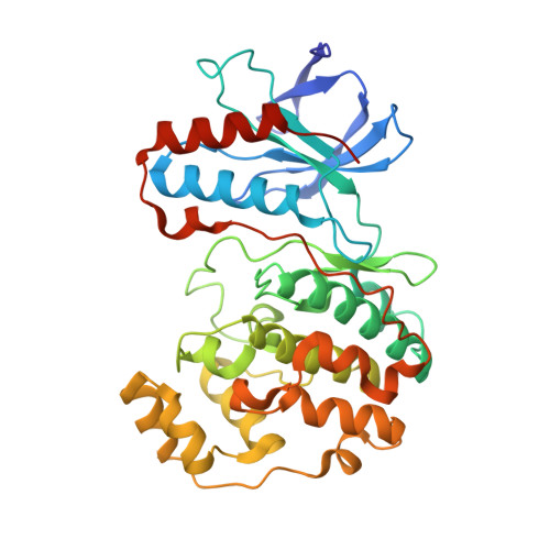
    4-Anilinoquinazolines represent an important class of protein kinase inhibitor. Modes of binding for two members of this inhibitor class were determined by X-ray crystallographic analysis of one inhibitor (4-[3-hydroxyanilino]-6,7-dimethoxyquinazoline) in complex with cyclin-dependent kinase 2 (CDK2) and the other (4-[3-methylsulfanylanilino]-6,7-dimethoxyquinazoline) in complex with p38 kinase. In both inhibitor/kinase structures, the 4-anilinoquinazoline was bound in the ATP site with the quinazoline ring system oriented along the peptide strand that links the two domains of the protein and with the anilino substituent projecting into a hydrophobic pocket within the protein interior. In each case, the nitrogen at position-1 of the quinazoline accepted a hydrogen bond from a backbone NH (CDK2, Leu-83; p38, Met-109) of the domain connector strand, and aromatic hydrogen atoms at C2 and C8 interacted with backbone carbonyl oxygen atoms of the peptide strand. The anilino group of the CDK2-bound compound was essentially coplanar with the quinazoline ring system and occupied a pocket between Lys-33 and Phe-80. For the p38-bound inhibitor, the anilino group was angled out of plane and was positioned between Lys-53 and Thr-106 in a manner similar to that observed for the aryl substituent of the pyridinylimidazole class of inhibitor.


  • Organizational Affiliation

    Glaxo Wellcome Inc., Five Moore Drive, Research Triangle Park, North Carolina 27709, USA.


Macromolecules
Find similar proteins by:  (by identity cutoff)  |  3D Structure
Entity ID: 1
MoleculeChains Sequence LengthOrganismDetailsImage
P38 KINASE360Homo sapiensMutation(s): 0 
EC: 2.7.1
UniProt & NIH Common Fund Data Resources
Find proteins for Q16539 (Homo sapiens)
Explore Q16539 
Go to UniProtKB:  Q16539
PHAROS:  Q16539
GTEx:  ENSG00000112062 
Entity Groups  
Sequence Clusters30% Identity50% Identity70% Identity90% Identity95% Identity100% Identity
UniProt GroupQ16539
Sequence Annotations
Expand
  • Reference Sequence
Small Molecules
Ligands 1 Unique
IDChains Name / Formula / InChI Key2D Diagram3D Interactions
MSQ
Query on MSQ

Download Ideal Coordinates CCD File 
B [auth A]4-[3-METHYLSULFANYLANILINO]-6,7-DIMETHOXYQUINAZOLINE
C17 H17 N3 O2 S
FUSDVOSGGMBSMK-UHFFFAOYSA-N
Binding Affinity Annotations 
IDSourceBinding Affinity
MSQ PDBBind:  1DI9 IC50: 5000 (nM) from 1 assay(s)
Experimental Data & Validation

Experimental Data

  • Method: X-RAY DIFFRACTION
  • Resolution: 2.60 Å
  • R-Value Free: 0.253 
  • R-Value Work: 0.209 
  • Space Group: P 21 21 21
Unit Cell:
Length ( Å )Angle ( ˚ )
a = 45.176α = 90
b = 84.862β = 90
c = 127.015γ = 90
Software Package:
Software NamePurpose
DENZOdata reduction
SCALEPACKdata scaling
CNSrefinement
CNSphasing

Structure Validation

View Full Validation Report


Ligand Structure Quality Assessment 


Entry History 

Deposition Data

Revision History  (Full details and data files)

  • Version 1.0: 2000-11-29
    Type: Initial release
  • Version 1.1: 2008-04-27
    Changes: Version format compliance
  • Version 1.2: 2011-07-13
    Changes: Version format compliance
  • Version 1.3: 2024-02-07
    Changes: Data collection, Database references, Derived calculations