1DHK

STRUCTURE OF PORCINE PANCREATIC ALPHA-AMYLASE


Experimental Data Snapshot

  • Method: X-RAY DIFFRACTION
  • Resolution: 1.85 Å
  • R-Value Free: 0.220 
  • R-Value Work: 0.183 
  • R-Value Observed: 0.183 

wwPDB Validation   3D Report Full Report


This is version 3.1 of the entry. See complete history


Literature

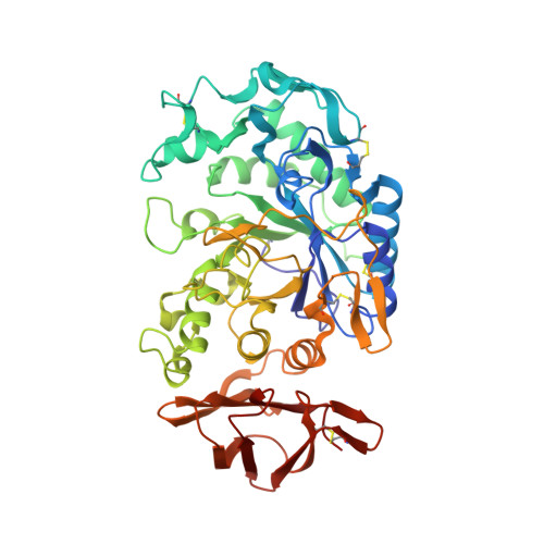
Substrate mimicry in the active center of a mammalian alpha-amylase: structural analysis of an enzyme-inhibitor complex.

Bompard-Gilles, C.Rousseau, P.Rouge, P.Payan, F.

(1996) Structure 4: 1441-1452

  • DOI: https://doi.org/10.1016/s0969-2126(96)00151-7
  • Primary Citation of Related Structures:  
    1DHK

  • PubMed Abstract: 

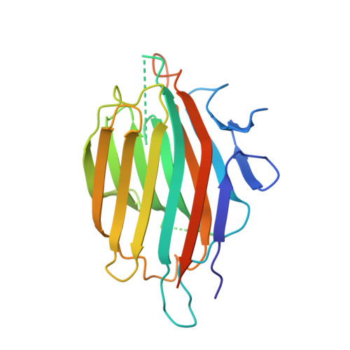
    alpha-Amylases catalyze the hydrolysis of glycosidic linkages in starch and other related polysaccharides. The alpha-amylase inhibitor (alpha-Al) from the bean Phaseolus vulgaris belongs to a family of plant defence proteins and is a potent inhibitor of mammalian alpha-amylases. The structure of pig pancreatic alpha-amylase (PPA) in complex with both a carbohydrate inhibitor (acarbose) and a proteinaceous inhibitor (Tendamistat) is known, but the catalytic mechanism is poorly understood. The crystal structure of pig pancreatic alpha-amylase complexed with alpha-Al was refined to 1.85 A resolution. It reveals that in complex with PPA, the inhibitor has the typical dimer structure common to legume lectins. Two hairpin loops extending out from the jellyroll fold of a monomer interact directly with the active site region of the enzyme molecule, with the inhibitor molecule filling the whole substrate-docking region of the PPA. The inhibitor makes substrate-mimetic interactions with binding subsites of the enzyme and targets catalytic residues in the active site. Binding of inhibitor induces structural changes at the active site of the enzyme. The present analysis reveals that there are extensive interactions between the inhibitor and residues that are highly conserved in the active site of alpha-amylases; alpha-Al1 inactivates PPA through elaborate blockage of substrate-binding sites. It provides a basis to design peptide analogue inhibitors. alpha-Amylase inhibition is of interest from several points of view, for example the treatment of diabetes and for crop protection.


  • Organizational Affiliation

    AFMB-IBSM-CNRS, Marseille, France.


Macromolecules
Find similar proteins by:  (by identity cutoff)  |  3D Structure
Entity ID: 1
MoleculeChains Sequence LengthOrganismDetailsImage
PORCINE PANCREATIC ALPHA-AMYLASE496Sus scrofaMutation(s): 0 
EC: 3.2.1.1
UniProt
Find proteins for P00690 (Sus scrofa)
Explore P00690 
Go to UniProtKB:  P00690
Entity Groups  
Sequence Clusters30% Identity50% Identity70% Identity90% Identity95% Identity100% Identity
UniProt GroupP00690
Sequence Annotations
Expand
  • Reference Sequence
Find similar proteins by:  (by identity cutoff)  |  3D Structure
Entity ID: 2
MoleculeChains Sequence LengthOrganismDetailsImage
BEAN LECTIN-LIKE INHIBITOR223Phaseolus vulgarisMutation(s): 0 
UniProt
Find proteins for P02873 (Phaseolus vulgaris)
Explore P02873 
Go to UniProtKB:  P02873
Entity Groups  
Sequence Clusters30% Identity50% Identity70% Identity90% Identity95% Identity100% Identity
UniProt GroupP02873
Sequence Annotations
Expand
  • Reference Sequence
Oligosaccharides

Help

Entity ID: 3
MoleculeChains Length2D Diagram Glycosylation3D Interactions
2-acetamido-2-deoxy-beta-D-glucopyranose-(1-4)-2-acetamido-2-deoxy-beta-D-glucopyranose
C, D
2N-Glycosylation
Glycosylation Resources
GlyTouCan:  G42666HT
GlyCosmos:  G42666HT
GlyGen:  G42666HT
Small Molecules
Modified Residues  1 Unique
IDChains TypeFormula2D DiagramParent
PCA
Query on PCA
A
L-PEPTIDE LINKINGC5 H7 N O3GLN
Experimental Data & Validation

Experimental Data

  • Method: X-RAY DIFFRACTION
  • Resolution: 1.85 Å
  • R-Value Free: 0.220 
  • R-Value Work: 0.183 
  • R-Value Observed: 0.183 
  • Space Group: C 1 2 1
Unit Cell:
Length ( Å )Angle ( ˚ )
a = 151.6α = 90
b = 79.4β = 91.54
c = 68γ = 90
Software Package:
Software NamePurpose
DENZOdata reduction
SCALEPACKdata scaling
AMoREphasing
X-PLORrefinement

Structure Validation

View Full Validation Report


Entry History 

Deposition Data

Revision History  (Full details and data files)

  • Version 1.0: 1997-12-24
    Type: Initial release
  • Version 1.1: 2008-03-24
    Changes: Version format compliance
  • Version 1.2: 2011-07-13
    Changes: Non-polymer description, Version format compliance
  • Version 2.0: 2019-12-25
    Changes: Data collection, Database references, Derived calculations, Polymer sequence
  • Version 3.0: 2020-07-29
    Type: Remediation
    Reason: Carbohydrate remediation
    Changes: Advisory, Atomic model, Data collection, Derived calculations, Structure summary
  • Version 3.1: 2024-04-03
    Changes: Data collection, Database references, Refinement description, Structure summary