1CRX

CRE RECOMBINASE/DNA COMPLEX REACTION INTERMEDIATE I


Experimental Data Snapshot

  • Method: X-RAY DIFFRACTION
  • Resolution: 2.40 Å
  • R-Value Free: 0.263 
  • R-Value Work: 0.201 

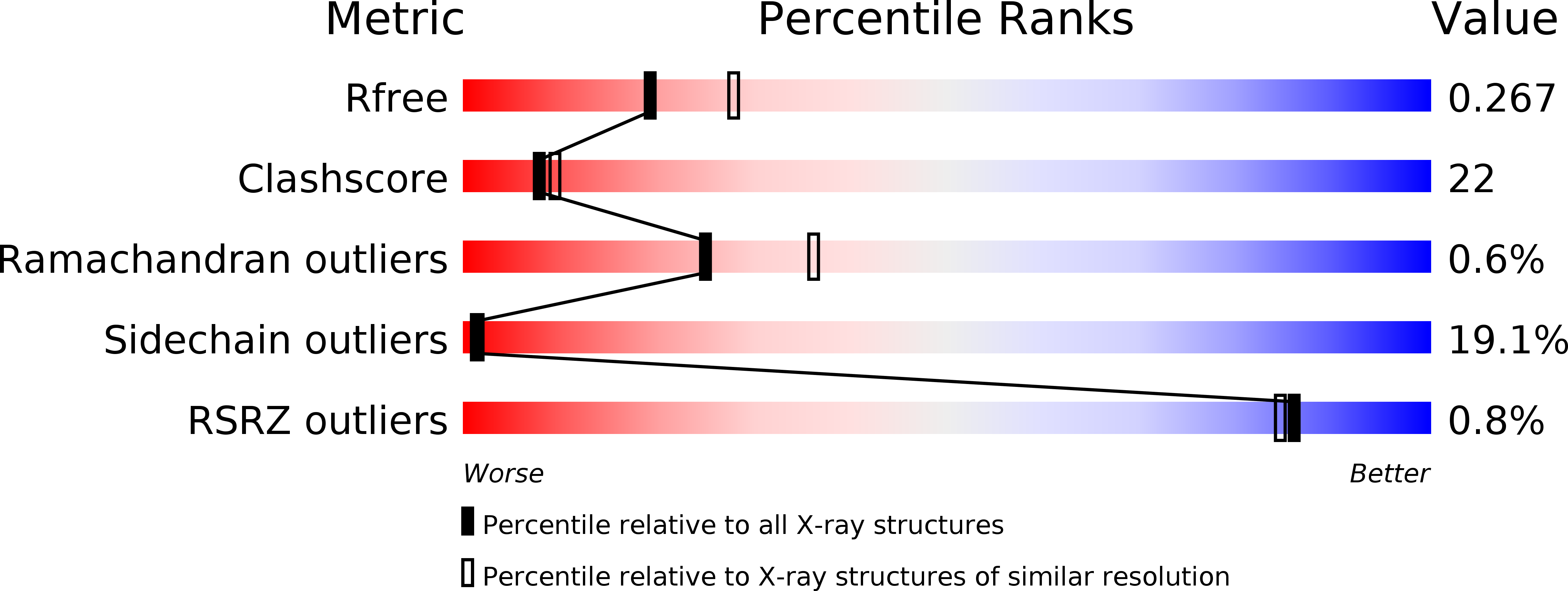
wwPDB Validation   3D Report Full Report


This is version 1.2 of the entry. See complete history


Literature

Structure of Cre recombinase complexed with DNA in a site-specific recombination synapse.

Guo, F.Gopaul, D.N.van Duyne, G.D.

(1997) Nature 389: 40-46

  • DOI: https://doi.org/10.1038/37925
  • Primary Citation of Related Structures:  
    1CRX

  • PubMed Abstract: 

    During site-specific DNA recombination, which brings about genetic rearrangement in processes such as viral integration and excision and chromosomal segregation, recombinase enzymes recognize specific DNA sequences and catalyse the reciprocal exchange of DNA strands between these sites. The bacteriophage recombinase Cre catalyses site-specific recombination between two 34-base-pair loxP sites. The crystal structure at 2.4 A resolution of Cre bound to a loxP substrate reveals an intermediate in the recombination reaction, in which a Cre molecule has cleaved the substrate to form a covalent 3'-phosphotyrosine linkage with the DNA. Four recombinases and two loxP sites form a synapsed structure in which the DNA resembles models of four-way Holliday-Junction intermediates. The Cre-loxP complex challenges models of site-specific recombination that require large changes in quaternary structure. Subtle allosteric changes at the carboxy termini of the Cre subunits may instead coordinate the cleavage and strand-exchange reactions.


  • Organizational Affiliation

    Johnson Research Foundation and Department of Biochemistry and Biophysics, University of Pennsylvania School of Medicine, Philadelphia 19104, USA.


Macromolecules

Find similar proteins by:  (by identity cutoff)  |  3D Structure
Entity ID: 4
MoleculeChains Sequence LengthOrganismDetailsImage
CRE RECOMBINASEE [auth A],
F [auth B]
322Punavirus P1Mutation(s): 0 
Gene Names: CRE
UniProt
Find proteins for P06956 (Escherichia phage P1)
Explore P06956 
Go to UniProtKB:  P06956
Entity Groups  
Sequence Clusters30% Identity50% Identity70% Identity90% Identity95% Identity100% Identity
UniProt GroupP06956
Sequence Annotations
Expand
  • Reference Sequence

Find similar nucleic acids by:  Sequence   |   3D Structure  

Entity ID: 1
MoleculeChains LengthOrganismImage
DNA (5'-D(*TP*AP*TP*AP*AP*CP*TP*TP*CP*GP*TP*AP*TP*AP*G)-3')A [auth C]15N/A
Sequence Annotations
Expand
  • Reference Sequence

Find similar nucleic acids by:  Sequence   |   3D Structure  

Entity ID: 2
MoleculeChains LengthOrganismImage
DNA (5'-D(*AP*TP*AP*TP*GP*CP*TP*AP*TP*AP*CP*GP*AP*AP*GP*TP*TP*AP*T)-3')B [auth D],
D [auth F]
19N/A
Sequence Annotations
Expand
  • Reference Sequence

Find similar nucleic acids by:  Sequence   |   3D Structure  

Entity ID: 3
MoleculeChains LengthOrganismImage
DNA (5'-D(P*AP*TP*AP*AP*CP*TP*TP*CP*GP*TP*AP*TP*AP*GP*C)-3')C [auth E]15N/A
Sequence Annotations
Expand
  • Reference Sequence
Small Molecules
Ligands 1 Unique
IDChains Name / Formula / InChI Key2D Diagram3D Interactions
PO4
Query on PO4

Download Ideal Coordinates CCD File 
G [auth B]PHOSPHATE ION
O4 P
NBIIXXVUZAFLBC-UHFFFAOYSA-K
Experimental Data & Validation

Experimental Data

  • Method: X-RAY DIFFRACTION
  • Resolution: 2.40 Å
  • R-Value Free: 0.263 
  • R-Value Work: 0.201 
  • Space Group: C 2 2 21
Unit Cell:
Length ( Å )Angle ( ˚ )
a = 107.7α = 90
b = 121β = 90
c = 180.4γ = 90
Software Package:
Software NamePurpose
MLPHAREphasing
LOCALmodel building
X-PLORrefinement
DENZOdata reduction
SCALEPACKdata scaling
LOCALphasing

Structure Validation

View Full Validation Report


Entry History 

Deposition Data

Revision History  (Full details and data files)

  • Version 1.0: 1998-10-14
    Type: Initial release
  • Version 1.1: 2008-05-22
    Changes: Version format compliance
  • Version 1.2: 2011-07-13
    Changes: Version format compliance