1BLN

ANTI-P-GLYCOPROTEIN FAB MRK-16


Experimental Data Snapshot

  • Method: X-RAY DIFFRACTION
  • Resolution: 2.80 Å
  • R-Value Free: 0.287 
  • R-Value Work: 0.209 
  • R-Value Observed: 0.209 

wwPDB Validation   3D Report Full Report


This is version 1.4 of the entry. See complete history


Literature

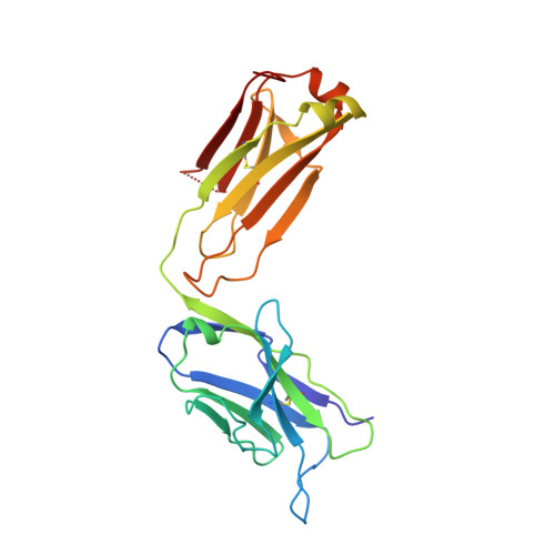
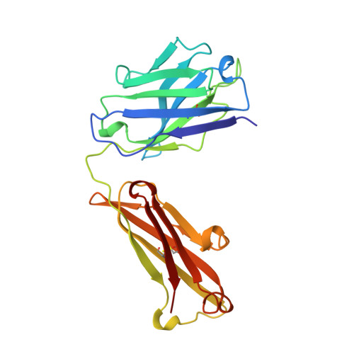
Mode of binding of anti-P-glycoprotein antibody MRK-16 to its antigen. A crystallographic and molecular modeling study.

Vasudevan, S.Tsuruo, T.Rose, D.R.

(1998) J Biol Chem 273: 25413-25419

  • DOI: https://doi.org/10.1074/jbc.273.39.25413
  • Primary Citation of Related Structures:  
    1BLN

  • PubMed Abstract: 

    Monoclonal antibody MRK-16 recognizes a discontinuous extracellular epitope on the multidrug resistance-associated ATP-binding cassette transporter, P-glycoprotein. The atomic basis for specificity of this antibody is of interest because of its potential as a modulator of P-glycoprotein activity. The crystal structure of Fab MRK-16 is reported to a resolution of 2.8 A. A structure for a portion of the epitope was derived by comparison to regions of solved structures with similar primary sequence. This has permitted a proposal for the mode of binding of the peptide epitope to the antibody, in which the peptide makes specific contacts with complementarity-determining regions H1, H2, and H3 from the heavy chain and L3 from the light chain. These interactions are consistent with epitope mapping studies and with the observation that MRK-16 is specific for human class I P-glycoprotein. This result identifies side chains in MRK-16 that would be amenable to alteration in antibody engineering experiments to derive improved multidrug resistance inhibitors for clinical use during chemotherapy. In particular, Arg-H97 contacts both Glu-746 and Asp-744 of the peptide, Arg-L96 contacts Asp-743, and Thr-H33 interacts with Thr-747. All of these epitope residues were implicated in mediating specificity by epitope mapping studies.


  • Organizational Affiliation

    Ontario Cancer Institute and Department of Medical Biophysics, University of Toronto, Toronto M5G 2M9, Canada.


Macromolecules
Find similar proteins by:  (by identity cutoff)  |  3D Structure
Entity ID: 1
MoleculeChains Sequence LengthOrganismDetailsImage
PROTEIN (MONOCLONAL ANTIBODY MRK-16 (LIGHT CHAIN))
A, C
214Mus musculusMutation(s): 0 
Entity Groups  
Sequence Clusters30% Identity50% Identity70% Identity90% Identity95% Identity100% Identity
Sequence Annotations
Expand
  • Reference Sequence
Find similar proteins by:  (by identity cutoff)  |  3D Structure
Entity ID: 2
MoleculeChains Sequence LengthOrganismDetailsImage
PROTEIN (MONOCLONAL ANTIBODY MRK-16 (HEAVY CHAIN))
B, D
218Mus musculusMutation(s): 0 
Entity Groups  
Sequence Clusters30% Identity50% Identity70% Identity90% Identity95% Identity100% Identity
Sequence Annotations
Expand
  • Reference Sequence
Experimental Data & Validation

Experimental Data

  • Method: X-RAY DIFFRACTION
  • Resolution: 2.80 Å
  • R-Value Free: 0.287 
  • R-Value Work: 0.209 
  • R-Value Observed: 0.209 
  • Space Group: P 1 21 1
Unit Cell:
Length ( Å )Angle ( ˚ )
a = 54.49α = 90
b = 67.774β = 97.61
c = 117.186γ = 90
Software Package:
Software NamePurpose
X-PLORmodel building
X-PLORrefinement
X-PLORphasing

Structure Validation

View Full Validation Report


Entry History 

Deposition Data

Revision History  (Full details and data files)

  • Version 1.0: 1998-10-28
    Type: Initial release
  • Version 1.1: 2008-04-27
    Changes: Version format compliance
  • Version 1.2: 2011-07-13
    Changes: Derived calculations, Version format compliance
  • Version 1.3: 2019-10-30
    Changes: Data collection, Derived calculations
  • Version 1.4: 2023-08-09
    Changes: Data collection, Database references, Refinement description