Binding domain of human parathyroid hormone receptor: from conformation to function.
Pellegrini, M., Bisello, A., Rosenblatt, M., Chorev, M., Mierke, D.F.(1998) Biochemistry 37: 12737-12743
- PubMed: 9737850
- DOI: https://doi.org/10.1021/bi981265h
- Primary Citation of Related Structures:
1BL1 - PubMed Abstract:
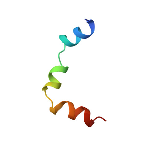
A 31 amino acid fragment of the extracellular N-terminus of the human G-protein coupled receptor for parathyroid hormone (PTH1R) has been structurally characterized by NMR and molecular dynamics simulations. The fragment PTH1R[168-198] includes residues 173-189, shown by photoaffinity cross-linking to be a contact domain with position 13 of parathyroid hormone (PTH). The structure of PTH1R[168-198], determined in a micellar solution of dodecylphosphocholine to mimic the membrane environment, consists of three alpha-helices, separated by a well-defined turn and a flexible region. The topological orientation of PTH1R[168-198] was determined from nitroxide-radical induced relaxation of NMR signals utilizing 5- and 16-doxylstearic acid. The C-terminal helix (residues 190-196), consisting of seven amino acids of the first transmembrane domain, is very hydrophobic and embedded in the lipid core. This helix is preceded by a well-defined turn, forming an approximate 90 degrees bend, placing the other helices (residues 169-176 and 180-189), both of which are amphipathic, on the surface of the micelle. In this orientation, many hydrophilic residues of the receptor, including Glu177, Arg179, Arg181, Glu182, Asp185, and Arg186, are projecting toward the solvent available to form complementary Coulombic interactions with the polar residues of the principal binding domain of the ligand (e.g., Arg25, Lys26, Lys27, Asp30, and His32). Given that the binding domain of PTH adopts an amphipathic alpha-helix which lies on the membrane, we visualize ligand binding as a two stage process involving a nonspecific hydrophobic interaction of amphipathic helices with the membrane, followed by two-dimensional diffusion leading to highly specific, ligand-receptor interaction.
Organizational Affiliation:
Department of Molecular Pharmacology, Physiology, & Biotechnology, Division of Biology & Medicine, Brown University, Providence, Rhode Island 02912, USA.