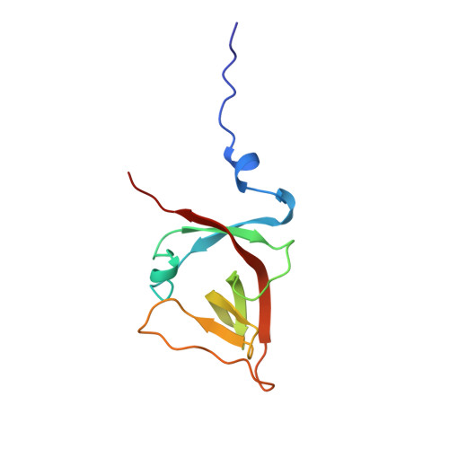
The UmuD' protein filament and its potential role in damage induced mutagenesis.
Peat, T.S., Frank, E.G., McDonald, J.P., Levine, A.S., Woodgate, R., Hendrickson, W.A.(1996) Structure 4: 1401-1412
- PubMed: 8994967
- DOI: https://doi.org/10.1016/s0969-2126(96)00148-7
- Primary Citation of Related Structures:
1AY9 - PubMed Abstract:
Damage induced 'SOS mutagenesis' may occur transiently as part of the global SOS response to DNA damage in bacteria. A key participant in this process is the UmuD protein, which is produced in an inactive from but converted to the active form, UmuD', by a RecA-mediated self-cleavage reaction. UmuD', together with UmuC and activated RecA (RecA*), enables the DNA polymerase III holoenzyme to replicate across chemical and UV induced lesions. The efficiency of this reaction depends on several intricate protein-protein interactions. Recent X-ray crystallographic analysis shows that in addition to forming molecular dimers, the N- and C-terminal tails of UmuD' extend from a globular beta structure to associate and produce crystallized filaments. We have investigated this phenomenon and find that these filaments appear to relate to biological activity. Higher order oligomers are found in solution with UmuD', but not with UmuD nor with a mutant of UmuD' lacking the extended N terminus. Deletion of the N terminus of UmuD' does not affect its ability to form molecular dimers but does severely compromise its ability to interact with a RecA-DNA filament and to participate in mutagenesis. Mutations in the C terminus of UmuD' result in both gain and loss of function for mutagenesis. The activation of UmuD to UmuD' appears to cause a large conformational change in the protein which allows it to form oligomers in solution at physiologically relevant concentrations. Properties of these oligomers are consistent with the filament structures seen in crystals of UmuD'.
Organizational Affiliation:
Department of Biochemistry and Molecular Biophysics, Columbia University, New York, NY 10032, USA.