1AY7

RIBONUCLEASE SA COMPLEX WITH BARSTAR


Experimental Data Snapshot

  • Method: X-RAY DIFFRACTION
  • Resolution: 1.70 Å
  • R-Value Free: 0.190 
  • R-Value Work: 0.160 
  • R-Value Observed: 0.162 

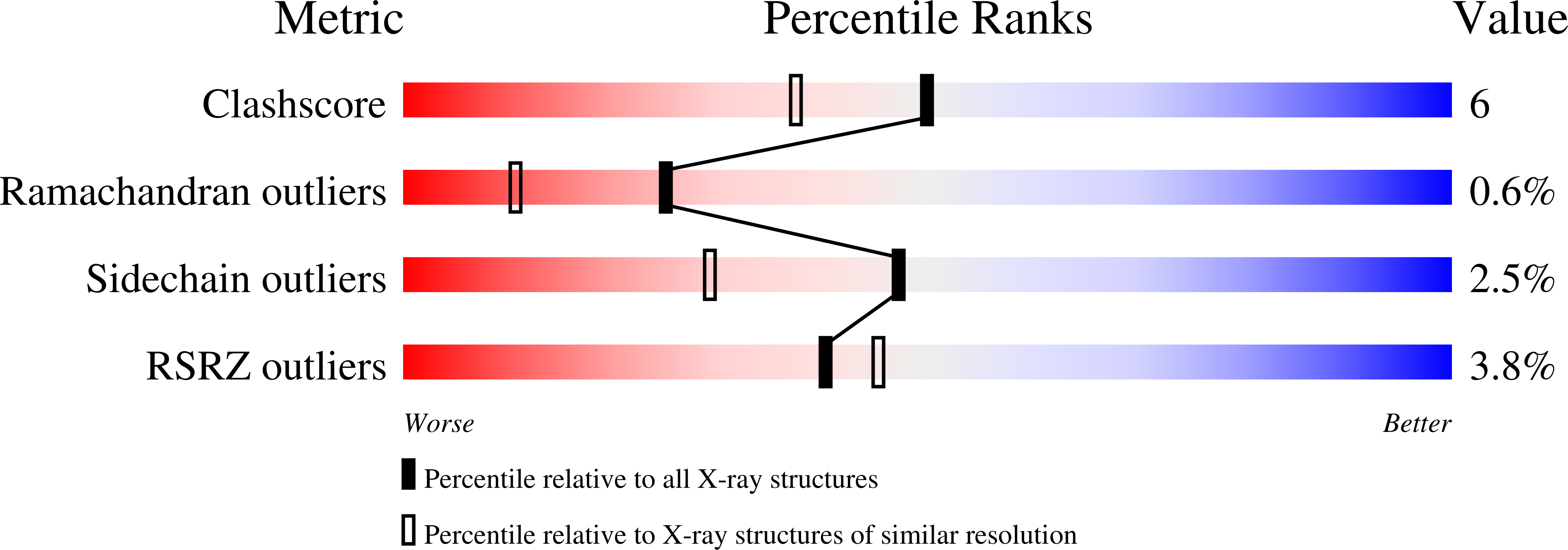
wwPDB Validation   3D Report Full Report


This is version 1.3 of the entry. See complete history


Literature

Recognition of RNase Sa by the inhibitor barstar: structure of the complex at 1.7 A resolution.

Sevcik, J.Urbanikova, L.Dauter, Z.Wilson, K.S.

(1998) Acta Crystallogr D Biol Crystallogr 54: 954-963

  • DOI: https://doi.org/10.1107/s0907444998004429
  • Primary Citation of Related Structures:  
    1AY7

  • PubMed Abstract: 

    We report the 1.7 A resolution structure of RNase Sa complexed with the polypeptide inhibitor barstar. The crystals are in the hexagonal space group P65 with unit-cell dimensions a = b = 56.9, c = 135.8 A and the asymmetric unit contains one molecule of the complex. RNase Sa is an extracellular microbial ribonuclease produced by Streptomyces aureofaciens. Barstar is the natural inhibitor of barnase, the ribonuclease of Bacillus amyloliquefaciens. It inhibits RNase Sa and barnase in a similar manner by steric blocking of the active site. The structure of RNase Sa is very similar to that observed in crystals of the native enzyme and its complexes with nucleotides. Barstar retains the structure found in its complex with barnase. The accessible surface area of protein buried in the complex is about 300 A2 smaller and there are fewer hydrogen bonds in the enzyme-inhibitor interface in RNase Sa-barstar than in barnase-barstar, providing an explanation of the reduced binding affinity in the former. Previous studies of barstar complexes have used mutants of the inhibitor and this is the first structure which includes wild-type barstar.


  • Organizational Affiliation

    Institute of Molecular Biology, Slovak Academy of Sciences, Dubravska cesta 21, 84251 Bratislava, Slovak Republic.


Macromolecules
Find similar proteins by:  (by identity cutoff)  |  3D Structure
Entity ID: 1
MoleculeChains Sequence LengthOrganismDetailsImage
GUANYL-SPECIFIC RIBONUCLEASE SA96Kitasatospora aureofaciensMutation(s): 0 
EC: 3.1.27.3
UniProt
Find proteins for P05798 (Kitasatospora aureofaciens)
Explore P05798 
Go to UniProtKB:  P05798
Entity Groups  
Sequence Clusters30% Identity50% Identity70% Identity90% Identity95% Identity100% Identity
UniProt GroupP05798
Sequence Annotations
Expand
  • Reference Sequence
Find similar proteins by:  (by identity cutoff)  |  3D Structure
Entity ID: 2
MoleculeChains Sequence LengthOrganismDetailsImage
BARSTAR89Bacillus amyloliquefaciensMutation(s): 0 
UniProt
Find proteins for P11540 (Bacillus amyloliquefaciens)
Explore P11540 
Go to UniProtKB:  P11540
Entity Groups  
Sequence Clusters30% Identity50% Identity70% Identity90% Identity95% Identity100% Identity
UniProt GroupP11540
Sequence Annotations
Expand
  • Reference Sequence
Experimental Data & Validation

Experimental Data

  • Method: X-RAY DIFFRACTION
  • Resolution: 1.70 Å
  • R-Value Free: 0.190 
  • R-Value Work: 0.160 
  • R-Value Observed: 0.162 
  • Space Group: P 65
Unit Cell:
Length ( Å )Angle ( ˚ )
a = 56.95α = 90
b = 56.95β = 90
c = 135.8γ = 120
Software Package:
Software NamePurpose
DENZOdata reduction
SCALEPACKdata scaling
AMoREphasing
REFMACrefinement

Structure Validation

View Full Validation Report


Entry History 

Deposition Data

Revision History  (Full details and data files)

  • Version 1.0: 1999-03-02
    Type: Initial release
  • Version 1.1: 2008-03-24
    Changes: Version format compliance
  • Version 1.2: 2011-07-13
    Changes: Version format compliance
  • Version 1.3: 2023-08-02
    Changes: Data collection, Database references, Refinement description