Kunitz-type soybean trypsin inhibitor revisited: refined structure of its complex with porcine trypsin reveals an insight into the interaction between a homologous inhibitor from Erythrina caffra and tissue-type plasminogen activator.
Song, H.K., Suh, S.W.(1998) J Mol Biol 275: 347-363
- PubMed: 9466914
- DOI: https://doi.org/10.1006/jmbi.1997.1469
- Primary Citation of Related Structures:
1AVU, 1AVW, 1AVX - PubMed Abstract:
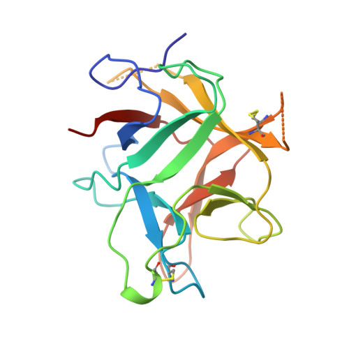
The Kunitz-type trypsin inhibitor from soybean (STI) consists of 181 amino acid residues with two disulfide bridges. Its crystal structures have been determined in complex with porcine pancreatic trypsin in two crystal forms (an orthorhombic form at 1.75 A resolution and a tetragonal form at 1.9 A) and in the free state at 2.3 A resolution. They have been refined to crystallographic R-values of 18.9%, 21.6% and 19.8%, respectively. The three models of STI reported here represent a significant improvement over the partial inhibitor structure in the complex, which was previously determined at a nominal resolution of 2.6 A by the multiple isomorphous replacement method. This study provides the first high-resolution picture of the complex between a Kunitz-type proteinase inhibitor with its cognate proteinase. Many of the external loops of STI show high B-factors, both in the free and the complexed states, except the reactive site loop whose B-factors are dramatically reduced upon complexation. The reactive site loop of STI adopts a canonical conformation similar to those in other substrate-like inhibitors. The P1 carbonyl group displays no out-of-plane displacement and thus retains a nominal trigonal planar geometry. Modeling studies on the complex between a homologous Kunitz-type trypsin inhibitor DE-3 from Erythrina caffra and the human tissue-type plasminogen activator reveal a new insight into the specific interactions which could play a crucial role in their binding.
Organizational Affiliation:
Department of Chemistry, College of Natural Sciences, Seoul National University, Korea.