1AQL

CRYSTAL STRUCTURE OF BOVINE BILE-SALT ACTIVATED LIPASE COMPLEXED WITH TAUROCHOLATE


Experimental Data Snapshot

  • Method: X-RAY DIFFRACTION
  • Resolution: 2.80 Å
  • R-Value Free: 0.275 
  • R-Value Work: 0.211 
  • R-Value Observed: 0.211 

wwPDB Validation   3D Report Full Report


This is version 1.6 of the entry. See complete history


Literature

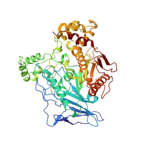
The crystal structure of bovine bile salt activated lipase: insights into the bile salt activation mechanism.

Wang, X.Wang, C.S.Tang, J.Dyda, F.Zhang, X.C.

(1997) Structure 5: 1209-1218

  • DOI: https://doi.org/10.1016/s0969-2126(97)00271-2
  • Primary Citation of Related Structures:  
    1AKN, 1AQL

  • PubMed Abstract: 

    The intestinally located pancreatic enzyme, bile salt activated lipase (BAL), possesses unique activities for digesting different kinds of lipids. It also differs from other lipases in a requirement of bile salts for activity. A structure-based explanation for these unique properties has not been reached so far due to the absence of a three-dimensional structure. The crystal structures of bovine BAL and its complex with taurocholate have been determined at 2.8 A resolution. The overall structure of BAL belongs to the alpha/beta hydrolase fold family. Two bile salt binding sites were found in each BAL molecule within the BAL-taurocholate complex structure. One of these sites is located close to a hairpin loop near the active site. Upon the binding of taurocholate, this loop becomes less mobile and assumes a different conformation. The other bile salt binding site is located remote from the active site. In both structures, BAL forms similar dimers with the active sites facing each other. Bile salts activate BAL by binding to a relatively short ten-residue loop near the active site, and stabilize the loop in an open conformation. Presumably, this conformational change leads to the formation of the substrate-binding site, as suggested from kinetic data. The BAL dimer observed in the crystal structure may also play a functional role under physiological conditions.


  • Organizational Affiliation

    Crystallography Program, Oklahoma Medical Research Foundation, Oklahoma City 73104, USA.


Macromolecules
Find similar proteins by:  (by identity cutoff)  |  3D Structure
Entity ID: 1
MoleculeChains Sequence LengthOrganismDetailsImage
BILE-SALT ACTIVATED LIPASE
A, B
532Bos taurusMutation(s): 0 
EC: 3.1.1.13
Membrane Entity: Yes 
UniProt
Find proteins for P30122 (Bos taurus)
Explore P30122 
Go to UniProtKB:  P30122
Entity Groups  
Sequence Clusters30% Identity50% Identity70% Identity90% Identity95% Identity100% Identity
UniProt GroupP30122
Sequence Annotations
Expand
  • Reference Sequence
Experimental Data & Validation

Experimental Data

  • Method: X-RAY DIFFRACTION
  • Resolution: 2.80 Å
  • R-Value Free: 0.275 
  • R-Value Work: 0.211 
  • R-Value Observed: 0.211 
  • Space Group: P 21 21 2
Unit Cell:
Length ( Å )Angle ( ˚ )
a = 130.23α = 90
b = 104.09β = 90
c = 120.18γ = 90
Software Package:
Software NamePurpose
SADIEdata collection
SAINTdata reduction
X-PLORmodel building
X-PLORrefinement
SADIEdata reduction
SAINTdata scaling
X-PLORphasing

Structure Validation

View Full Validation Report


Entry History 

Deposition Data

Revision History  (Full details and data files)

  • Version 1.0: 1998-08-05
    Type: Initial release
  • Version 1.1: 2008-03-24
    Changes: Version format compliance
  • Version 1.2: 2011-07-13
    Changes: Non-polymer description, Version format compliance
  • Version 1.3: 2011-08-24
    Changes: Other
  • Version 1.4: 2018-03-07
    Changes: Data collection, Other
  • Version 1.5: 2020-07-29
    Type: Remediation
    Reason: Carbohydrate remediation
    Changes: Advisory, Data collection, Derived calculations, Structure summary
  • Version 1.6: 2023-08-02
    Changes: Database references, Refinement description, Structure summary