1A4R

G12V MUTANT OF HUMAN PLACENTAL CDC42 GTPASE IN THE GDP FORM


Experimental Data Snapshot

  • Method: X-RAY DIFFRACTION
  • Resolution: 2.50 Å
  • R-Value Free: 0.280 
  • R-Value Work: 0.228 
  • R-Value Observed: 0.228 

wwPDB Validation   3D Report Full Report


Ligand Structure Quality Assessment 


This is version 1.4 of the entry. See complete history


Literature

Nucleotide binding to the G12V-mutant of Cdc42 investigated by X-ray diffraction and fluorescence spectroscopy: two different nucleotide states in one crystal.

Rudolph, M.G.Wittinghofer, A.Vetter, I.R.

(1999) Protein Sci 8: 778-787

  • DOI: https://doi.org/10.1110/ps.8.4.778
  • Primary Citation of Related Structures:  
    1A4R

  • PubMed Abstract: 

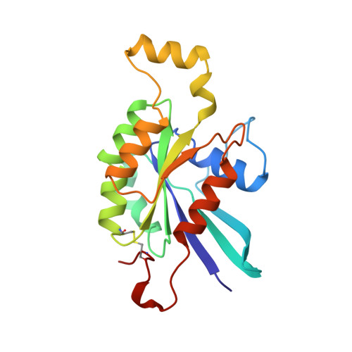
    The 2.5 A crystal structure of the full length human placental isoform of the Gly12 to Val mutant Cdc42 protein (Cdc42(G12V)) bound to both GDP/Mg2+ and GDPNH2 (guanosine-5'-diphospho-beta-amidate) is reported. The crystal contains two molecules in the asymmetric unit, of which one has bound GDP/Mg2+, while the other has bound GDPNH2 without a Mg2+ ion. Crystallization of the protein was induced via hydrolysis of the Cdc42 x GppNHp complex by the presence of contaminating alkaline phosphatase activity in combination with the crystallization conditions. This prompted us to compare the binding characteristics of GDPNH2 vs. GDP. The amino group of GDPNH2 drastically reduces the affinity to Cdc42 in comparison with that of GDP, causes the loss of the Mg2+ ion, and apparently also increases the conformational flexibility of the protein as seen in the crystal. Both the switch I and switch II regions are visible in the electron density of the GDP-bound molecule, but not in the molecule bound to GDPNH2. The C-terminus containing the CaaX-motif is partly ordered in both molecules due to an intramolecular disulfide bond formed between Cys105/Cys188 and Cys305/Cys388, respectively.


  • Organizational Affiliation

    Max-Planck Institut für molekulare Physiologie, Abteilung Strukturelle Biologie, Dortmund, Germany.


Macromolecules
Find similar proteins by:  (by identity cutoff)  |  3D Structure
Entity ID: 1
MoleculeChains Sequence LengthOrganismDetailsImage
G25K GTP-BINDING PROTEIN
A, B
191Homo sapiensMutation(s): 1 
UniProt & NIH Common Fund Data Resources
Find proteins for P60953 (Homo sapiens)
Explore P60953 
Go to UniProtKB:  P60953
PHAROS:  P60953
GTEx:  ENSG00000070831 
Entity Groups  
Sequence Clusters30% Identity50% Identity70% Identity90% Identity95% Identity100% Identity
UniProt GroupP60953
Sequence Annotations
Expand
  • Reference Sequence
Small Molecules
Binding Affinity Annotations 
IDSourceBinding Affinity
GNH PDBBind:  1A4R Kd: 220 (nM) from 1 assay(s)
Binding MOAD:  1A4R Kd: 220 (nM) from 1 assay(s)
GDP Binding MOAD:  1A4R Kd: 9.40e-3 (nM) from 1 assay(s)
Experimental Data & Validation

Experimental Data

  • Method: X-RAY DIFFRACTION
  • Resolution: 2.50 Å
  • R-Value Free: 0.280 
  • R-Value Work: 0.228 
  • R-Value Observed: 0.228 
  • Space Group: P 41 21 2
Unit Cell:
Length ( Å )Angle ( ˚ )
a = 98.65α = 90
b = 98.65β = 90
c = 104.16γ = 90
Software Package:
Software NamePurpose
DENZOdata reduction
SCALEPACKdata scaling
X-PLORmodel building
X-PLORrefinement
X-PLORphasing

Structure Validation

View Full Validation Report


Ligand Structure Quality Assessment 


Entry History 

Deposition Data

Revision History  (Full details and data files)

  • Version 1.0: 1999-03-02
    Type: Initial release
  • Version 1.1: 2008-03-24
    Changes: Version format compliance
  • Version 1.2: 2011-07-13
    Changes: Version format compliance
  • Version 1.3: 2021-11-03
    Changes: Data collection, Database references, Derived calculations
  • Version 1.4: 2023-08-02
    Changes: Refinement description