1ZSF

Crystal Structure of Complex of a Hydroxyethylamine Inhibitor with HIV-1 Protease at 2.0A Resolution


Experimental Data Snapshot

  • Method: X-RAY DIFFRACTION
  • Resolution: 1.98 Å
  • R-Value Free: 0.276 
  • R-Value Work: 0.221 
  • R-Value Observed: 0.222 

wwPDB Validation   3D Report Full Report


Ligand Structure Quality Assessment 


This is version 1.5 of the entry. See complete history


Literature

On the role of the R configuration of the reaction-intermediate isostere in HIV-1 protease-inhibitor binding: X-ray structure at 2.0 A resolution.

Duskova, J.Dohnalek, J.Skalova, T.Petrokova, H.Vondrackova, E.Hradilek, M.Konvalinka, J.Soucek, M.Brynda, J.Fabry, M.Sedlacek, J.Hasek, J.

(2006) Acta Crystallogr D Biol Crystallogr 62: 489-497

  • DOI: https://doi.org/10.1107/S0907444906006718
  • Primary Citation of Related Structures:  
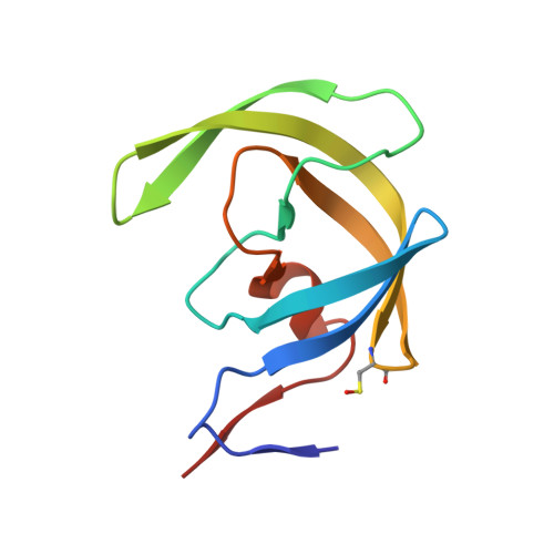
    1ZSF, 1ZSR

  • PubMed Abstract: 

    Peptidomimetic inhibitors of human immunodeficiency virus-1 protease are successful lead substances for the development of virostatic drugs against HIV as the causative agent of acquired immunodeficiency syndrome (AIDS). The hydroxyethylamine isostere of the proteolytic cleavage intermediate provides a suitable replacement for the peptide bond. A series of acyclic pseudopeptide inhibitors with the hydroxyethylamine isostere varying in chiral carbon configuration and P'2 residue type were structurally analysed by single-crystal X-ray crystallography. The compounds inhibit HIV protease with subnanomolar inhibition constants and block viral replication in tissue cultures. Here, the structure of such a complex with the R configuration of the isosteric group (PDB code 1zsf) is presented together with newly available synchrotron data for a complex with the S stereoisomer of the inhibitor (PDB code 1zsr). Comparison of the structure and binding with other complexes of HIV-1 protease and similar inhibitors contributes to the understanding of how these molecules bind to the wild-type form of this enzyme. The hydroxy group of the R stereoisomer interacts with one of the catalytic aspartic acids by a short hydrogen bond with rather extreme geometry. The change of configuration of the chiral carbon bearing the hydroxyl from S to R does not influence the inhibition efficiency in this case.


  • Organizational Affiliation

    Institute of Macromolecular Chemistry, Academy of Sciences of the Czech Republic, Heyrovského nám. 2, 162 06 Praha 6, Czech Republic. duskova@imc.cas.cz


Macromolecules
Find similar proteins by:  (by identity cutoff)  |  3D Structure
Entity ID: 1
MoleculeChains Sequence LengthOrganismDetailsImage
PROTEASE RETROPEPSIN
A, B
99Human immunodeficiency virus 1Mutation(s): 0 
EC: 3.4.23.16
UniProt
Find proteins for P03366 (Human immunodeficiency virus type 1 group M subtype B (isolate BH10))
Explore P03366 
Go to UniProtKB:  P03366
Entity Groups  
Sequence Clusters30% Identity50% Identity70% Identity90% Identity95% Identity100% Identity
UniProt GroupP03366
Sequence Annotations
Expand
  • Reference Sequence
Small Molecules
Ligands 1 Unique
IDChains Name / Formula / InChI Key2D Diagram3D Interactions
0ZS
Query on 0ZS

Download Ideal Coordinates CCD File 
C [auth B]N-{(2R,3S)-3-[(tert-butoxycarbonyl)amino]-2-hydroxy-4-phenylbutyl}-L-phenylalanyl-L-alpha-glutamyl-L-phenylalaninamide
C38 H49 N5 O8
MPMUDVMRFYJRLP-QEUNAIBPSA-N
Modified Residues  1 Unique
IDChains TypeFormula2D DiagramParent
CSO
Query on CSO
A, B
L-PEPTIDE LINKINGC3 H7 N O3 SCYS
Binding Affinity Annotations 
IDSourceBinding Affinity
0ZS Binding MOAD:  1ZSF Ki: 0.12 (nM) from 1 assay(s)
Biologically Interesting Molecules (External Reference) 1 Unique
Experimental Data & Validation

Experimental Data

Unit Cell:
Length ( Å )Angle ( ˚ )
a = 62.864α = 90
b = 62.864β = 90
c = 82.375γ = 120
Software Package:
Software NamePurpose
DENZOdata reduction
SCALEPACKdata scaling
AMoREphasing
SHELXL-97refinement

Structure Validation

View Full Validation Report


Ligand Structure Quality Assessment 


Entry History 

Deposition Data

Revision History  (Full details and data files)

  • Version 1.0: 2006-04-25
    Type: Initial release
  • Version 1.1: 2007-10-16
    Changes: Version format compliance
  • Version 1.2: 2011-07-13
    Changes: Atomic model, Database references, Derived calculations, Non-polymer description, Structure summary, Version format compliance
  • Version 1.3: 2012-12-12
    Changes: Other
  • Version 1.4: 2023-08-23
    Changes: Data collection, Database references, Derived calculations, Refinement description
  • Version 1.5: 2023-11-15
    Changes: Data collection