1ZS9

Crystal structure of human enolase-phosphatase E1


Experimental Data Snapshot

  • Method: X-RAY DIFFRACTION
  • Resolution: 1.70 Å
  • R-Value Free: 0.241 
  • R-Value Work: 0.208 
  • R-Value Observed: 0.208 

wwPDB Validation   3D Report Full Report


This is version 1.2 of the entry. See complete history


Literature

Crystal Structure of Human E1 Enzyme and its Complex with a Substrate Analog Reveals the Mechanism of its Phosphatase/Enolase

Wang, H.Pang, H.Bartlam, M.Rao, Z.

(2005) J Mol Biol 348: 917-926

  • DOI: https://doi.org/10.1016/j.jmb.2005.01.072
  • Primary Citation of Related Structures:  
    1YNS, 1ZS9

  • PubMed Abstract: 

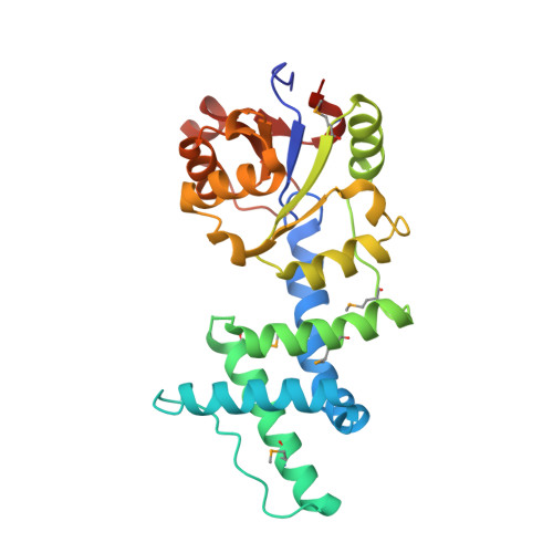
    Enolase-phosphatase E1 (MASA) is a bifunctional enzyme in the ubiquitous methionine salvage pathway that catalyzes the continuous reactions of 2,3-diketo-5-methylthio-1-phosphopentane to yield the aci-reductone metabolite using Mg2+ as cofactor. In this study, we have determined the crystal structure of MASA and its complex with a substrate analog to 1.7A resolution by multi-wavelength anomalous diffraction and molecular replacement techniques, respectively. The structures support the proposed mechanism of phosphatase activity and further suggest the probable mechanism of enolization. We establish a model for substrate binding to describe in detail the enzymatic reaction and the formation of the transition state, which will provide insight into the reaction mechanisms of other enzymes in the same family.


  • Organizational Affiliation

    Laboratory of Structural Biology, School of Medicine, Tsinghua University, Beijing 100084, China.


Macromolecules
Find similar proteins by:  (by identity cutoff)  |  3D Structure
Entity ID: 1
MoleculeChains Sequence LengthOrganismDetailsImage
E-1 ENZYME261Homo sapiensMutation(s): 6 
UniProt & NIH Common Fund Data Resources
Find proteins for Q9UHY7 (Homo sapiens)
Explore Q9UHY7 
Go to UniProtKB:  Q9UHY7
PHAROS:  Q9UHY7
GTEx:  ENSG00000145293 
Entity Groups  
Sequence Clusters30% Identity50% Identity70% Identity90% Identity95% Identity100% Identity
UniProt GroupQ9UHY7
Sequence Annotations
Expand
  • Reference Sequence
Small Molecules
Modified Residues  1 Unique
IDChains TypeFormula2D DiagramParent
MSE
Query on MSE
A
L-PEPTIDE LINKINGC5 H11 N O2 SeMET
Experimental Data & Validation

Experimental Data

  • Method: X-RAY DIFFRACTION
  • Resolution: 1.70 Å
  • R-Value Free: 0.241 
  • R-Value Work: 0.208 
  • R-Value Observed: 0.208 
  • Space Group: P 21 21 21
Unit Cell:
Length ( Å )Angle ( ˚ )
a = 54.017α = 90
b = 57.546β = 90
c = 87.318γ = 90
Software Package:
Software NamePurpose
AUTOMARdata collection
AUTOMARdata reduction
SOLVEphasing
CNSrefinement

Structure Validation

View Full Validation Report


Entry History 

Deposition Data

Revision History  (Full details and data files)

  • Version 1.0: 2005-06-21
    Type: Initial release
  • Version 1.1: 2008-04-30
    Changes: Version format compliance
  • Version 1.2: 2011-07-13
    Changes: Version format compliance