1ZPQ

STRUCTURE OF BACTERIOPHAGE LAMBDA CII protein


Experimental Data Snapshot

  • Method: X-RAY DIFFRACTION
  • Resolution: 2.80 Å
  • R-Value Free: 0.286 
  • R-Value Work: 0.255 
  • R-Value Observed: 0.257 

wwPDB Validation   3D Report Full Report


This is version 1.4 of the entry. See complete history


Literature

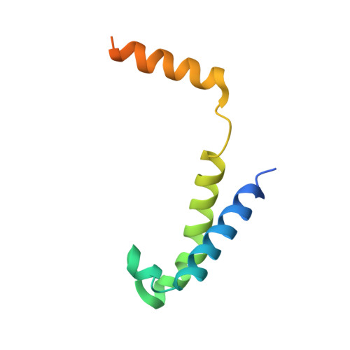
Crystal Structure of Bacteriophage lambdacII and Its DNA Complex.

Jain, D.Kim, Y.Maxwell, K.L.Beasley, S.Zhang, R.Gussin, G.N.Edwards, A.M.Darst, S.A.

(2005) Mol Cell 19: 259-269

  • DOI: https://doi.org/10.1016/j.molcel.2005.06.006
  • Primary Citation of Related Structures:  
    1ZPQ, 1ZS4

  • PubMed Abstract: 

    The tetrameric cII protein from bacteriophage lambda activates transcription from the phage promoters P(RE), P(I), and P(AQ) by binding to two direct repeats that flank the promoter -35 element. Here, we present the X-ray crystal structure of cII alone (2.8 A resolution) and in complex with its DNA operator from P(RE) (1.7 A resolution). The structures provide a basis for modeling of the activation complex with the RNA polymerase holoenzyme, and point to the key role for the RNA polymerase alpha subunit C-terminal domain (alphaCTD) in cII-dependent activation, which forms a bridge of protein/protein interactions between cII and the RNA polymerase sigma subunit. The model makes specific predictions for protein/protein interactions between cII and alphaCTD, and between alphaCTD and sigma, which are supported by previous genetic studies.


  • Organizational Affiliation

    The Rockefeller University, 1230 York Avenue, New York, New York 10021, USA.


Macromolecules
Find similar proteins by:  (by identity cutoff)  |  3D Structure
Entity ID: 1
MoleculeChains Sequence LengthOrganismDetailsImage
Regulatory protein CII
A, B, C, D
97Lambdavirus lambdaMutation(s): 0 
Gene Names: CII
UniProt
Find proteins for P03042 (Escherichia phage lambda)
Explore P03042 
Go to UniProtKB:  P03042
Entity Groups  
Sequence Clusters30% Identity50% Identity70% Identity90% Identity95% Identity100% Identity
UniProt GroupP03042
Sequence Annotations
Expand
  • Reference Sequence
Experimental Data & Validation

Experimental Data

  • Method: X-RAY DIFFRACTION
  • Resolution: 2.80 Å
  • R-Value Free: 0.286 
  • R-Value Work: 0.255 
  • R-Value Observed: 0.257 
  • Space Group: C 2 2 21
Unit Cell:
Length ( Å )Angle ( ˚ )
a = 59.56α = 90
b = 108.16β = 90
c = 111.45γ = 90
Software Package:
Software NamePurpose
HKL-2000data collection
SCALEPACKdata scaling
SOLVEphasing
REFMACrefinement
HKL-2000data reduction

Structure Validation

View Full Validation Report


Entry History 

Revision History  (Full details and data files)

  • Version 1.0: 2005-08-23
    Type: Initial release
  • Version 1.1: 2008-04-30
    Changes: Version format compliance
  • Version 1.2: 2011-07-13
    Changes: Version format compliance
  • Version 1.3: 2011-10-05
    Changes: Structure summary
  • Version 1.4: 2024-02-14
    Changes: Data collection, Database references