1ZD3

Human soluble epoxide hydrolase 4-(3-cyclohexyluriedo)-butyric acid complex


Experimental Data Snapshot

  • Method: X-RAY DIFFRACTION
  • Resolution: 2.30 Å
  • R-Value Free: 0.254 
  • R-Value Work: 0.212 

wwPDB Validation   3D Report Full Report


Ligand Structure Quality Assessment 


This is version 1.3 of the entry. See complete history


Literature

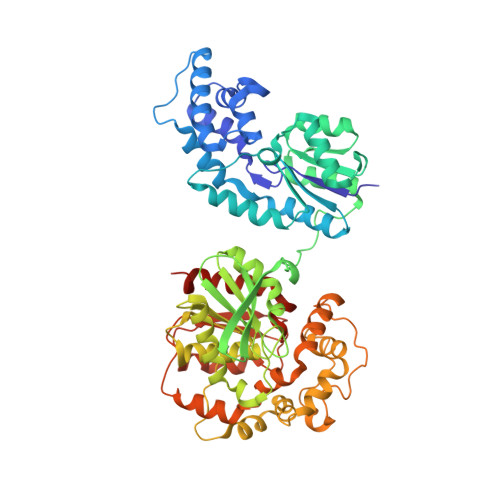
Human soluble epoxide hydrolase: structural basis of inhibition by 4-(3-cyclohexylureido)-carboxylic acids

Gomez, G.A.Morisseau, C.Hammock, B.D.Christianson, D.W.

(2006) Protein Sci 15: 58-64

  • DOI: https://doi.org/10.1110/ps.051720206
  • Primary Citation of Related Structures:  
    1ZD2, 1ZD3, 1ZD4, 1ZD5

  • PubMed Abstract: 

    X-ray crystal structures of human soluble epoxide hydrolase (sEH) complexed with four different dialkylurea inhibitors bearing pendant carboxylate "tails" of varying length have been determined at 2.3-3.0 A resolution. Similarities among inhibitor binding modes reinforce the proposed roles of Y381 and/or Y465 as general acids that protonate the epoxide ring of the substrate in concert with nucleophilic attack of D333 at the electrophilic epoxide carbon. Additionally, the binding of these inhibitors allows us to model the binding mode of the endogenous substrate 14,15-epoxyeicosatrienoic acid. Contrasts among inhibitor binding modes include opposite orientations of inhibitor binding in the active-site hydrophobic tunnel. Alternative binding orientations observed for this series of inhibitors to human sEH, as well as the binding of certain dialkylurea inhibitors to human sEH and murine sEH, complicate the structure-based design of human sEH inhibitors with potential pharmaceutical applications in the treatment of hypertension. Thus, with regard to the optimization of inhibitor designs targeting human sEH, it is critical that human sEH and not murine sEH be utilized for inhibitor screening, and it is critical that structures of human sEH-inhibitor complexes be determined to verify inhibitor binding orientations that correlate with measured affinities.


  • Organizational Affiliation

    Department of Chemistry, University of Pennsylvania, Philadelphia, PA 19104-6323, USA.


Macromolecules
Find similar proteins by:  (by identity cutoff)  |  3D Structure
Entity ID: 1
MoleculeChains Sequence LengthOrganismDetailsImage
epoxide hydrolase 2, cytoplasmic555Homo sapiensMutation(s): 0 
Gene Names: EPHX2
EC: 3.3.2.3
UniProt & NIH Common Fund Data Resources
Find proteins for P34913 (Homo sapiens)
Explore P34913 
Go to UniProtKB:  P34913
PHAROS:  P34913
GTEx:  ENSG00000120915 
Entity Groups  
Sequence Clusters30% Identity50% Identity70% Identity90% Identity95% Identity100% Identity
UniProt GroupP34913
Sequence Annotations
Expand
  • Reference Sequence
Small Molecules
Binding Affinity Annotations 
IDSourceBinding Affinity
NC4 PDBBind:  1ZD3 IC50: 5.00e+5 (nM) from 1 assay(s)
BindingDB:  1ZD3 IC50: 3.60e+4 (nM) from 1 assay(s)
Binding MOAD:  1ZD3 IC50: 5.00e+5 (nM) from 1 assay(s)
Experimental Data & Validation

Experimental Data

  • Method: X-RAY DIFFRACTION
  • Resolution: 2.30 Å
  • R-Value Free: 0.254 
  • R-Value Work: 0.212 
  • Space Group: P 65 2 2
Unit Cell:
Length ( Å )Angle ( ˚ )
a = 92.41α = 90
b = 92.41β = 90
c = 243.316γ = 120
Software Package:
Software NamePurpose
HKL-2000data collection
SCALEPACKdata scaling
AMoREphasing
CNSrefinement
HKL-2000data reduction

Structure Validation

View Full Validation Report


Ligand Structure Quality Assessment 


Entry History 

Deposition Data

Revision History  (Full details and data files)

  • Version 1.0: 2006-03-07
    Type: Initial release
  • Version 1.1: 2008-04-30
    Changes: Version format compliance
  • Version 1.2: 2011-07-13
    Changes: Version format compliance
  • Version 1.3: 2023-08-23
    Changes: Data collection, Database references, Derived calculations, Refinement description