1ZBA

Foot-and-Mouth Disease virus serotype A1061 complexed with oligosaccharide receptor.


Experimental Data Snapshot

  • Method: X-RAY DIFFRACTION
  • Resolution: 2.00 Å
  • R-Value Observed: 0.183 

wwPDB Validation   3D Report Full Report


This is version 2.2 of the entry. See complete history


Literature

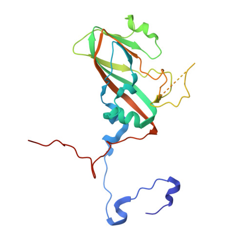
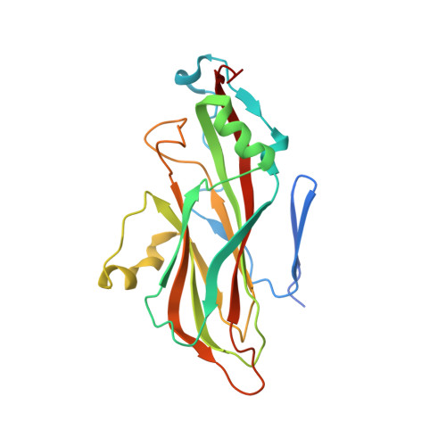
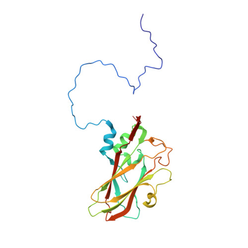
Structure of Foot-and-mouth disease virus serotype A1061 alone and complexed with oligosaccharide receptor: receptor conservation in the face of antigenic variation.

Fry, E.E.Newman, J.W.Curry, S.Najjam, S.Jackson, T.Blakemore, W.Lea, S.M.Miller, L.Burman, A.King, A.M.Stuart, D.I.

(2005) J Gen Virol 86: 1909-1920

  • DOI: https://doi.org/10.1099/vir.0.80730-0
  • Primary Citation of Related Structures:  
    1ZBA, 1ZBE

  • PubMed Abstract: 

    Foot-and-mouth disease viruses (FMDVs) target epithelial cells via integrin receptors, but can acquire the capacity to bind cell-surface heparan sulphate (or alternative receptors) on passage in cell culture. Vaccine viruses must be propagated in cell culture and, hence, some rationale for the selection of variants in this process is important. Crystal structures are available for type O, A and C viruses and also for a complex of type O strain O(1)BFS with heparin. The structure of FMDV A10(61) (a cell culture-adapted strain) complexed with heparin has now been determined. This virus has an RGSD motif in place of the otherwise conserved RGD integrin-binding motif and the potential to bind heparan sulphate (suggested by sequence analyses). FMDV A10(61) was closely similar in structure to other serotypes, deviating most in antigenic sites. The VP1 GH loop comprising the integrin-binding motif was disordered. Heparin bound at a similar site and in a similar conformation to that seen in the analogous complex with O(1)BFS, although the binding had a lower affinity and was more ionic.


  • Organizational Affiliation

    Division of Structural Biology, The Henry Wellcome Building for Genomic Medicine, Roosevelt Drive, Headington, Oxford OX3 7BN, UK.


Macromolecules
Find similar proteins by:  (by identity cutoff)  |  3D Structure
Entity ID: 1
MoleculeChains Sequence LengthOrganismDetailsImage
Coat protein VP1A [auth 1]212Foot-and-mouth disease virusMutation(s): 0 
UniProt
Find proteins for P03306 (Foot-and-mouth disease virus (strain A10-61))
Explore P03306 
Go to UniProtKB:  P03306
Entity Groups  
Sequence Clusters30% Identity50% Identity70% Identity90% Identity95% Identity100% Identity
UniProt GroupP03306
Sequence Annotations
Expand
  • Reference Sequence
Find similar proteins by:  (by identity cutoff)  |  3D Structure
Entity ID: 2
MoleculeChains Sequence LengthOrganismDetailsImage
Coat protein VP2B [auth 2]218Foot-and-mouth disease virusMutation(s): 1 
UniProt
Find proteins for P03306 (Foot-and-mouth disease virus (strain A10-61))
Explore P03306 
Go to UniProtKB:  P03306
Entity Groups  
Sequence Clusters30% Identity50% Identity70% Identity90% Identity95% Identity100% Identity
UniProt GroupP03306
Sequence Annotations
Expand
  • Reference Sequence
Find similar proteins by:  (by identity cutoff)  |  3D Structure
Entity ID: 3
MoleculeChains Sequence LengthOrganismDetailsImage
Coat protein VP3C [auth 3]221Foot-and-mouth disease virusMutation(s): 0 
UniProt
Find proteins for P03306 (Foot-and-mouth disease virus (strain A10-61))
Explore P03306 
Go to UniProtKB:  P03306
Entity Groups  
Sequence Clusters30% Identity50% Identity70% Identity90% Identity95% Identity100% Identity
UniProt GroupP03306
Sequence Annotations
Expand
  • Reference Sequence
Find similar proteins by:  (by identity cutoff)  |  3D Structure
Entity ID: 4
MoleculeChains Sequence LengthOrganismDetailsImage
Coat protein VP4D [auth 4]85Foot-and-mouth disease virusMutation(s): 0 
UniProt
Find proteins for P03306 (Foot-and-mouth disease virus (strain A10-61))
Explore P03306 
Go to UniProtKB:  P03306
Entity Groups  
Sequence Clusters30% Identity50% Identity70% Identity90% Identity95% Identity100% Identity
UniProt GroupP03306
Sequence Annotations
Expand
  • Reference Sequence
Oligosaccharides

Help

Entity ID: 5
MoleculeChains Length2D Diagram Glycosylation3D Interactions
2-deoxy-6-O-sulfo-2-(sulfoamino)-alpha-D-glucopyranose-(1-4)-2-O-sulfo-alpha-L-idopyranuronic acid-(1-4)-2-deoxy-6-O-sulfo-2-(sulfoamino)-alpha-D-glucopyranoseE [auth A]3N/A
Glycosylation Resources
GlyTouCan:  G66470SO
GlyCosmos:  G66470SO
Experimental Data & Validation

Experimental Data

  • Method: X-RAY DIFFRACTION
  • Resolution: 2.00 Å
  • R-Value Observed: 0.183 
  • Space Group: H 3
Unit Cell:
Length ( Å )Angle ( ˚ )
a = 307α = 90
b = 307β = 90
c = 715γ = 120
Software Package:
Software NamePurpose
X-PLORrefinement

Structure Validation

View Full Validation Report


Entry History 

Deposition Data

Revision History  (Full details and data files)

  • Version 1.0: 2005-06-28
    Type: Initial release
  • Version 1.1: 2008-04-30
    Changes: Version format compliance
  • Version 1.2: 2011-07-13
    Changes: Non-polymer description, Version format compliance
  • Version 2.0: 2020-07-29
    Type: Remediation
    Reason: Carbohydrate remediation
    Changes: Atomic model, Data collection, Derived calculations, Structure summary
  • Version 2.1: 2021-10-20
    Changes: Database references, Structure summary
  • Version 2.2: 2024-02-14
    Changes: Data collection, Derived calculations