1Y9D

Pyruvate Oxidase variant V265A from Lactobacillus plantarum


Experimental Data Snapshot

  • Method: X-RAY DIFFRACTION
  • Resolution: 2.20 Å
  • R-Value Free: 0.238 
  • R-Value Work: 0.178 
  • R-Value Observed: 0.181 

wwPDB Validation   3D Report Full Report


Ligand Structure Quality Assessment 


This is version 1.4 of the entry. See complete history


Literature

The role of Val-265 for Flavin Adenine Dinulceotide (FAD) binding in pyruvate oxidase: FTIR, kinetic and crystallographic studies on the enzyme variant V265A

Wille, G.Ritter, M.Weiss, M.S.Konig, S.Mantele, W.Hubner, G.

(2005) Biochemistry 44: 5086-5094

  • DOI: https://doi.org/10.1021/bi047337o
  • Primary Citation of Related Structures:  
    1Y9D

  • PubMed Abstract: 

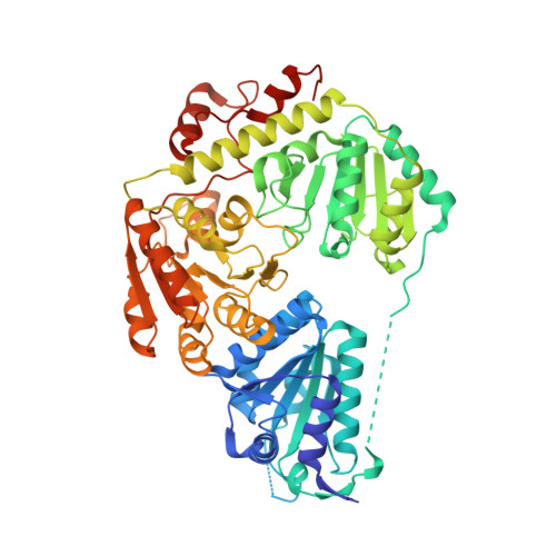
    In pyruvate oxidase (POX) from Lactobacillus plantarum, valine 265 participates in binding the cofactor FAD and is responsible for the strained conformation of its isoalloxazine moiety that is visible in the crystal structure of POX. The contrasting effects of the conservative amino acid exchange V265A on the enzyme's catalytic properties, cofactor affinity, and protein structure were investigated. The most prominent effect of the exchange was observed in the 2.2 A crystal structure of the mutant POX. While the overall structures of the wild-type and the variant are similar, flavin binding in particular is clearly different. Local disorder at the isoalloxazine binding site prevents modeling of the complete FAD cofactor and two protein loops of the binding site. Only the ADP moiety shows well-defined electron density, indicating an "anchor" function for this part of the molecule. This notion is corroborated by competition experiments where ADP was used to displace FAD from the variant enzyme. Despite the fact that the affinity of FAD binding in the variant is reduced, the catalytic properties are very similar to the wild-type, and the redox potential of the bound flavin is the same for both proteins. The rate of electron transfer toward the flavin during turnover is reduced to one-third compared to the wild-type, but k(cat) remains unchanged. Redox-triggered FTIR difference spectroscopy of free FAD shows the nu(C(10a)=N(1)) band at 1548 cm(-)(1). In POX-V265A, this band is found at 1538 cm(-)(1) and thus shifted less strongly than in wild-type POX where it is found at 1534 cm(-)(1). Taking these observations together, the conservative exchange V265A in POX has a surprisingly small effect on the catalytic properties of the enzyme, whereas the effect on the three-dimensional structure is rather big.


  • Organizational Affiliation

    Institut für Biochemie, Martin-Luther-Universität Halle-Wittenberg, Kurt-Mothes-Strasse 3, 06120 Halle, Germany. georg@bc.biochemtech.uni-halle.de


Macromolecules
Find similar proteins by:  (by identity cutoff)  |  3D Structure
Entity ID: 1
MoleculeChains Sequence LengthOrganismDetailsImage
Pyruvate oxidase
A, B, C, D
603Lactiplantibacillus plantarumMutation(s): 1 
Gene Names: pox5
EC: 1.2.3.3
UniProt
Find proteins for P37063 (Lactiplantibacillus plantarum (strain ATCC BAA-793 / NCIMB 8826 / WCFS1))
Explore P37063 
Go to UniProtKB:  P37063
Entity Groups  
Sequence Clusters30% Identity50% Identity70% Identity90% Identity95% Identity100% Identity
UniProt GroupP37063
Sequence Annotations
Expand
  • Reference Sequence
Small Molecules
Ligands 5 Unique
IDChains Name / Formula / InChI Key2D Diagram3D Interactions
FAD
Query on FAD

Download Ideal Coordinates CCD File 
I [auth A],
N [auth B],
R [auth C],
V [auth D]
FLAVIN-ADENINE DINUCLEOTIDE
C27 H33 N9 O15 P2
VWWQXMAJTJZDQX-UYBVJOGSSA-N
TPP
Query on TPP

Download Ideal Coordinates CCD File 
H [auth A],
M [auth B],
Q [auth C],
U [auth D]
THIAMINE DIPHOSPHATE
C12 H19 N4 O7 P2 S
AYEKOFBPNLCAJY-UHFFFAOYSA-O
SO4
Query on SO4

Download Ideal Coordinates CCD File 
G [auth A],
L [auth B],
O [auth C],
S [auth D]
SULFATE ION
O4 S
QAOWNCQODCNURD-UHFFFAOYSA-L
MG
Query on MG

Download Ideal Coordinates CCD File 
E [auth A],
J [auth B],
P [auth C],
T [auth D]
MAGNESIUM ION
Mg
JLVVSXFLKOJNIY-UHFFFAOYSA-N
NA
Query on NA

Download Ideal Coordinates CCD File 
F [auth A],
K [auth B]
SODIUM ION
Na
FKNQFGJONOIPTF-UHFFFAOYSA-N
Experimental Data & Validation

Experimental Data

  • Method: X-RAY DIFFRACTION
  • Resolution: 2.20 Å
  • R-Value Free: 0.238 
  • R-Value Work: 0.178 
  • R-Value Observed: 0.181 
  • Space Group: P 1 21 1
Unit Cell:
Length ( Å )Angle ( ˚ )
a = 94.66α = 90
b = 155.78β = 92.92
c = 100.75γ = 90
Software Package:
Software NamePurpose
REFMACrefinement
DENZOdata reduction
SCALEPACKdata scaling
MOLREPphasing

Structure Validation

View Full Validation Report


Ligand Structure Quality Assessment 


Entry History 

Deposition Data

Revision History  (Full details and data files)

  • Version 1.0: 2005-04-05
    Type: Initial release
  • Version 1.1: 2008-04-30
    Changes: Version format compliance
  • Version 1.2: 2011-07-13
    Changes: Version format compliance
  • Version 1.3: 2021-10-20
    Changes: Data collection, Database references, Derived calculations
  • Version 1.4: 2023-08-23
    Changes: Data collection, Refinement description