1XXR

Structure of a mannose-specific jacalin-related lectin from Morus Nigra in complex with mannose


Experimental Data Snapshot

  • Method: X-RAY DIFFRACTION
  • Resolution: 2.00 Å
  • R-Value Free: 0.211 
  • R-Value Work: 0.191 
  • R-Value Observed: 0.191 

wwPDB Validation   3D Report Full Report


Ligand Structure Quality Assessment 


This is version 1.4 of the entry. See complete history


Literature

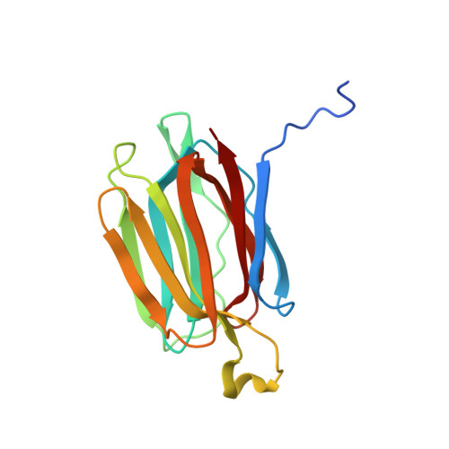
Structural analysis of the jacalin-related lectin MornigaM from the black mulberry (Morus nigra) in complex with mannose

Rabijns, A.Barre, A.Van Damme, E.J.M.Peumans, W.J.De Ranter, C.J.Rouge, P.

(2005) FEBS J 272: 3725-3732

  • DOI: https://doi.org/10.1111/j.1742-4658.2005.04801.x
  • Primary Citation of Related Structures:  
    1XXQ, 1XXR

  • PubMed Abstract: 

    The structures of MornigaM and the MornigaM-mannose complex have been determined at 1.8 A and 2.0 A resolution, respectively. Both structures adopt the typical beta-prism motif found in other jacalin-related lectins and their tetrameric assembly closely resembles that of jacalin. The carbohydrate-binding cavity of MornigaM readily binds mannose. No major structural rearrangements can be observed in MornigaM upon binding of mannose. These results allow corroboration of the structure-function relationships within the small group of Moraceae lectins.


  • Organizational Affiliation

    Laboratory of Analytical Chemistry and Medicinal Physicochemistry, Faculty of Pharmaceutical Sciences, K. U. Leuven, Belgium.


Macromolecules
Find similar proteins by:  (by identity cutoff)  |  3D Structure
Entity ID: 1
MoleculeChains Sequence LengthOrganismDetailsImage
mannose-binding lectin
A, B, C, D
161Morus nigraMutation(s): 0 
UniProt
Find proteins for Q8LGR3 (Morus nigra)
Explore Q8LGR3 
Go to UniProtKB:  Q8LGR3
Entity Groups  
Sequence Clusters30% Identity50% Identity70% Identity90% Identity95% Identity100% Identity
UniProt GroupQ8LGR3
Sequence Annotations
Expand
  • Reference Sequence
Small Molecules
Ligands 4 Unique
IDChains Name / Formula / InChI Key2D Diagram3D Interactions
MAN
Query on MAN

Download Ideal Coordinates CCD File 
G [auth B],
I [auth C],
K [auth D]
alpha-D-mannopyranose
C6 H12 O6
WQZGKKKJIJFFOK-PQMKYFCFSA-N
SO4
Query on SO4

Download Ideal Coordinates CCD File 
H [auth B]SULFATE ION
O4 S
QAOWNCQODCNURD-UHFFFAOYSA-L
GOL
Query on GOL

Download Ideal Coordinates CCD File 
F [auth A]GLYCEROL
C3 H8 O3
PEDCQBHIVMGVHV-UHFFFAOYSA-N
ACY
Query on ACY

Download Ideal Coordinates CCD File 
E [auth A],
J [auth C],
L [auth D]
ACETIC ACID
C2 H4 O2
QTBSBXVTEAMEQO-UHFFFAOYSA-N
Experimental Data & Validation

Experimental Data

  • Method: X-RAY DIFFRACTION
  • Resolution: 2.00 Å
  • R-Value Free: 0.211 
  • R-Value Work: 0.191 
  • R-Value Observed: 0.191 
  • Space Group: P 65
Unit Cell:
Length ( Å )Angle ( ˚ )
a = 110.908α = 90
b = 110.908β = 90
c = 160.021γ = 120
Software Package:
Software NamePurpose
CNSrefinement
DENZOdata reduction
SCALEPACKdata scaling
X-PLORphasing

Structure Validation

View Full Validation Report


Ligand Structure Quality Assessment 


Entry History 

Deposition Data

Revision History  (Full details and data files)

  • Version 1.0: 2005-10-25
    Type: Initial release
  • Version 1.1: 2008-04-30
    Changes: Version format compliance
  • Version 1.2: 2011-07-13
    Changes: Non-polymer description, Version format compliance
  • Version 1.3: 2020-07-29
    Type: Remediation
    Reason: Carbohydrate remediation
    Changes: Data collection, Derived calculations, Structure summary
  • Version 1.4: 2024-03-13
    Changes: Data collection, Database references, Structure summary