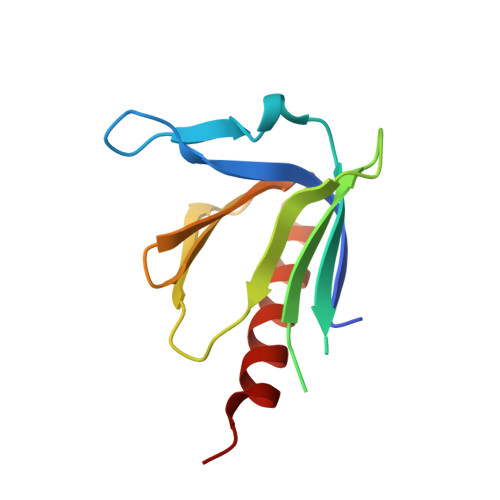
1XOD

Crystal structure of X. tropicalis Spred1 EVH-1 domain


Experimental Data Snapshot

  • Method: X-RAY DIFFRACTION
  • Resolution: 1.15 Å
  • R-Value Free: 0.177 
  • R-Value Work: 0.154 
  • R-Value Observed: 0.155 

wwPDB Validation   3D Report Full Report


This is version 1.3 of the entry. See complete history


Literature

1.15A Crystal structure of the X. tropicalis Spred1 EVH1 domain suggests a fourth distinct peptide-binding mechanism within the EVH1 family

Harmer, N.J.Sivak, J.M.Amaya, E.Blundell, T.L.

(2005) FEBS Lett 579: 1161-1166

  • DOI: https://doi.org/10.1016/j.febslet.2004.11.114
  • Primary Citation of Related Structures:  
    1XOD

  • PubMed Abstract: 

    The recently described Spred protein family has been implicated in the modulation of receptor tyrosine kinase signalling. We report the crystal structure of the Enabled/vasodilator-stimulated phosphoprotein homology-1 (EVH1) domain from Xenopus tropicalis Spred1, solved to 1.15 A resolution. This structure confirms that the Spred EVH1 adopts the pleckstrin-homology fold, with a similar secondary structure to Enabled. A translation of one of the peptide-binding groove beta-strands narrows this groove, whilst one end of the groove shows structural flexibility. We propose that Spred1 will bind peptides that are less proline-rich than other EVH1 domains, with conformational changes indicating an induced fit.


  • Organizational Affiliation

    Department of Biochemistry, 80 Tennis Court Road, Cambridge, CB2 1GA, UK. nic@cryst.bioc.cam.ac.uk


Macromolecules
Find similar proteins by:  (by identity cutoff)  |  3D Structure
Entity ID: 1
MoleculeChains Sequence LengthOrganismDetailsImage
Spred1
A, B
118Xenopus tropicalisMutation(s): 0 
Gene Names: Spred1
UniProt
Find proteins for Q66JG9 (Xenopus tropicalis)
Explore Q66JG9 
Go to UniProtKB:  Q66JG9
Entity Groups  
Sequence Clusters30% Identity50% Identity70% Identity90% Identity95% Identity100% Identity
UniProt GroupQ66JG9
Sequence Annotations
Expand
  • Reference Sequence
Small Molecules
Ligands 1 Unique
IDChains Name / Formula / InChI Key2D Diagram3D Interactions
GOL
Query on GOL

Download Ideal Coordinates CCD File 
C [auth B]GLYCEROL
C3 H8 O3
PEDCQBHIVMGVHV-UHFFFAOYSA-N
Experimental Data & Validation

Experimental Data

  • Method: X-RAY DIFFRACTION
  • Resolution: 1.15 Å
  • R-Value Free: 0.177 
  • R-Value Work: 0.154 
  • R-Value Observed: 0.155 
  • Space Group: P 1 21 1
Unit Cell:
Length ( Å )Angle ( ˚ )
a = 32.811α = 90
b = 38.193β = 95.68
c = 79.907γ = 90
Software Package:
Software NamePurpose
REFMACrefinement
HKL-2000data reduction
SCALEPACKdata scaling
AMoREphasing

Structure Validation

View Full Validation Report


Entry History 

Deposition Data

Revision History  (Full details and data files)

  • Version 1.0: 2005-01-25
    Type: Initial release
  • Version 1.1: 2008-04-30
    Changes: Version format compliance
  • Version 1.2: 2011-07-13
    Changes: Non-polymer description, Version format compliance
  • Version 1.3: 2023-08-23
    Changes: Data collection, Database references, Derived calculations, Refinement description