Structural Studies of a Two-domain Chitinase from Streptomyces griseus HUT6037
Kezuka, Y., Ohishi, M., Itoh, Y., Watanabe, J., Mitsutomi, M., Watanabe, T., Nonaka, T.(2006) J Mol Biol 358: 472-484
- PubMed: 16516924
- DOI: https://doi.org/10.1016/j.jmb.2006.02.013
- Primary Citation of Related Structures:
1WVU, 1WVV, 2DBT - PubMed Abstract:
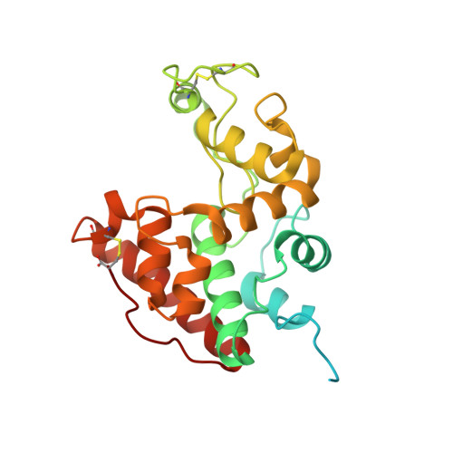
Chitinase C (ChiC) from Streptomyces griseus HUT6037 was the first glycoside hydrolase family 19 chitinase that was found in an organism other than higher plants. An N-terminal chitin-binding domain and a C-terminal catalytic domain connected by a linker peptide constitute ChiC. We determined the crystal structure of full-length ChiC, which is the only representative of the two-domain chitinases in the family. The catalytic domain has an alpha-helix-rich fold with a deep cleft containing a catalytic site, and lacks three loops on the domain surface compared with the catalytic domain of plant chitinases. The chitin-binding domain is an all-beta protein with two tryptophan residues (Trp59 and Trp60) aligned on the surface. We suggest the binding mechanism of tri-N-acetylchitotriose onto the chitin-binding domain on the basis of molecular dynamics (MD) simulations. In this mechanism, the ligand molecule binds well on the surface-exposed binding site through two stacking interactions and two hydrogen bonds and only Trp59 and Trp60 are involved in the binding. Furthermore, the flexibility of the Trp60 side-chain, which may be involved in adjusting the binding surface to fit the surface of crystalline chitin by the rotation of chi2 angle, is shown.
Organizational Affiliation:
Department of BioEngineering, Nagaoka University of Technology, Nagaoka, Niigata 940-2188, Japan.