1WU3

Crystal structure of recombinant murine interferon beta


Experimental Data Snapshot

  • Method: X-RAY DIFFRACTION
  • Resolution: 2.15 Å
  • R-Value Free: 0.247 
  • R-Value Work: 0.192 
  • R-Value Observed: 0.197 

wwPDB Validation   3D Report Full Report


This is version 1.4 of the entry. See complete history


Literature

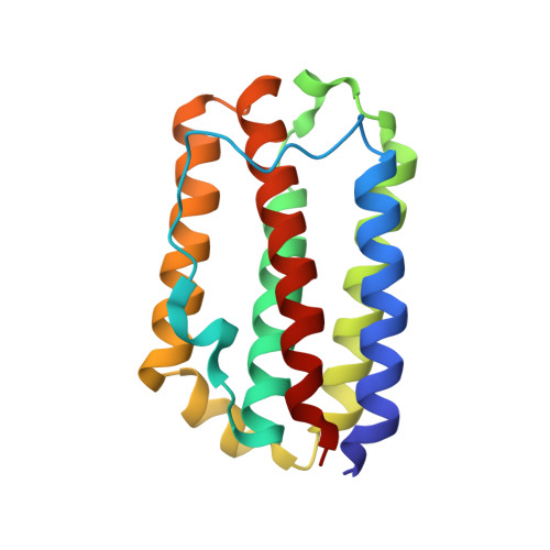
Refined crystal structure of recombinant murine interferon-beta at 2.15 A resolution

Senda, T.Saitoh, S.Mitsui, Y.

(1995) J Mol Biol 253: 187-207

  • DOI: https://doi.org/10.1006/jmbi.1995.0544
  • Primary Citation of Related Structures:  
    1WU3

  • PubMed Abstract: 

    The crystal structure of recombinant murine interferon-beta (reMuIFN-beta) has been refined at 2.15 A resolution using newly collected synchrotron data. Based on 11,228 reflections (8.0 to 2.15 A), a final R-factor of 19.1% (with a free R-factor of 25.8%) was obtained with a model obeying standard geometry within 0.013 A in bond lengths and 1.4 degrees in bond angles. Compared with the previously reported model, several amino acid residues in helix A are frame-shifted, the conformations are changed for parts of loops AB and BC, helix C is extended and a new short helix exists in loop CD. Evolutionary considerations taken together, the type I interferons appear to share common structural features with respect to the chain-folding topology and the hydrogen-bond networks between various polypeptide segments. Specifically, the disposition of the C-terminal segment of loop AB (after Arg33), known to be an important receptor-binding site, seems to be strictly maintained among the type I interferons. The exposed amino acid residues on helices A and C, which have recently been implicated as the binding site for another receptor molecule, are less well conserved. This may be responsible for varied cellular effects among the subtypes of type I interferons.


  • Organizational Affiliation

    Department of BioEngineering, Nagaoka University of Technology, Niigata, Japan.


Macromolecules
Find similar proteins by:  (by identity cutoff)  |  3D Structure
Entity ID: 1
MoleculeChains Sequence LengthOrganismDetailsImage
Interferon betaA [auth I]161Mus musculusMutation(s): 0 
UniProt
Find proteins for P01575 (Mus musculus)
Explore P01575 
Go to UniProtKB:  P01575
Entity Groups  
Sequence Clusters30% Identity50% Identity70% Identity90% Identity95% Identity100% Identity
UniProt GroupP01575
Sequence Annotations
Expand
  • Reference Sequence
Experimental Data & Validation

Experimental Data

  • Method: X-RAY DIFFRACTION
  • Resolution: 2.15 Å
  • R-Value Free: 0.247 
  • R-Value Work: 0.192 
  • R-Value Observed: 0.197 
  • Space Group: P 65
Unit Cell:
Length ( Å )Angle ( ˚ )
a = 71.4α = 90
b = 71.4β = 90
c = 79.6γ = 120
Software Package:
Software NamePurpose
REFMACrefinement
WEISdata reduction
SCALEdata scaling
PROTEINphasing

Structure Validation

View Full Validation Report


Entry History 

Deposition Data

Revision History  (Full details and data files)

  • Version 1.0: 2004-12-14
    Type: Initial release
  • Version 1.1: 2008-04-30
    Changes: Version format compliance
  • Version 1.2: 2011-07-13
    Changes: Version format compliance
  • Version 1.3: 2017-10-11
    Changes: Advisory
  • Version 1.4: 2024-03-13
    Changes: Advisory, Data collection, Database references