Crystal structures of Nipah and Hendra virus fusion core proteins
Lou, Z., Xu, Y., Xiang, K., Su, N., Qin, L., Li, X., Gao, G.F., Bartlam, M., Rao, Z.(2006) FEBS J 273: 4538-4547
- PubMed: 16972940
- DOI: https://doi.org/10.1111/j.1742-4658.2006.05459.x
- Primary Citation of Related Structures:
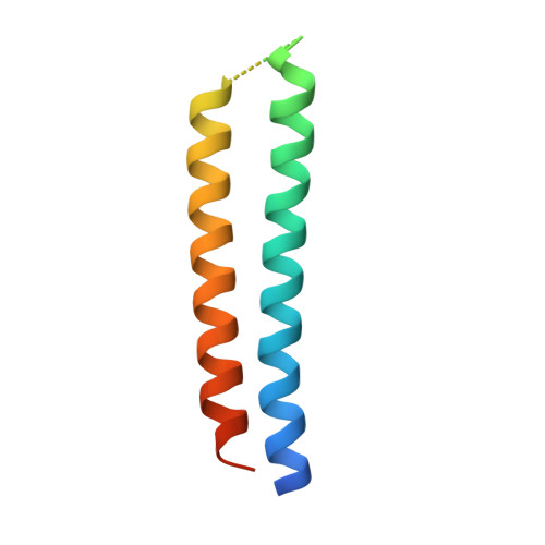
1WP8 - PubMed Abstract:
The Nipah and Hendra viruses are highly pathogenic paramyxoviruses that recently emerged from flying foxes to cause serious disease outbreaks in humans and livestock in Australia, Malaysia, Singapore and Bangladesh. Their unique genetic constitution, high virulence and wide host range set them apart from other paramyxoviruses. These characteristics have led to their classification into the new genus Henpavirus within the family Paramyxoviridae and to their designation as Biosafety Level 4 pathogens. The fusion protein, an enveloped glycoprotein essential for viral entry, belongs to the family of class I fusion proteins and is characterized by the presence of two heptad repeat (HR) regions, HR1 and HR2. These two regions associate to form a fusion-active hairpin conformation that juxtaposes the viral and cellular membranes to facilitate membrane fusion and enable subsequent viral entry. The Hendra and Nipah virus fusion core proteins were crystallized and their structures determined to 2.2 A resolution. The Nipah and Hendra fusion core structures are six-helix bundles with three HR2 helices packed against the hydrophobic grooves on the surface of a central coiled coil formed by three parallel HR1 helices in an oblique antiparallel manner. Because of the high level of conservation in core regions, it is proposed that the Nipah and Hendra virus fusion cores can provide a model for membrane fusion in all paramyxoviruses. The relatively deep grooves on the surface of the central coiled coil represent a good target site for drug discovery strategies aimed at inhibiting viral entry by blocking hairpin formation.
Organizational Affiliation:
Tsinghua-Nankai-IBP Joint Research Group for Structural Biology, Tsinghua University, Beijing, China.