


Crystal structure of a hypothetical protein from thermus thermophilus HB8
Agari, Y., Yokoyama, S., Kuramitsu, S.To be published.
Experimental Data Snapshot
wwPDB Validation 3D Report Full Report

Entity ID: 1 | |||||
|---|---|---|---|---|---|
| Molecule | Chains | Sequence Length | Organism | Details | Image |
| HYPOTHETICAL PROTEIN | 121 | Thermus thermophilus | Mutation(s): 0 | 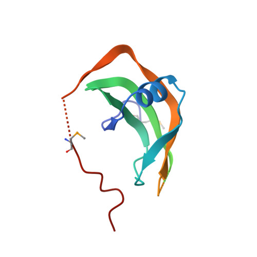 | |
UniProt | |||||
Find proteins for Q5SM30 (Thermus thermophilus (strain ATCC 27634 / DSM 579 / HB8)) Explore Q5SM30 Go to UniProtKB: Q5SM30 | |||||
Entity Groups | |||||
| Sequence Clusters | 30% Identity50% Identity70% Identity90% Identity95% Identity100% Identity | ||||
| UniProt Group | Q5SM30 | ||||
Sequence AnnotationsExpand | |||||
| |||||
| Modified Residues 1 Unique | |||||
|---|---|---|---|---|---|
| ID | Chains | Type | Formula | 2D Diagram | Parent |
| MSE Query on MSE | A | L-PEPTIDE LINKING | C5 H11 N O2 Se |  | MET |
| Length ( Å ) | Angle ( ˚ ) |
|---|---|
| a = 52.583 | α = 90 |
| b = 55.846 | β = 96.6 |
| c = 43.186 | γ = 90 |
| Software Name | Purpose |
|---|---|
| CNS | refinement |
| HKL-2000 | data reduction |
| SCALEPACK | data scaling |
| SOLVE | phasing |
RCSB PDB (citation) is hosted by
RCSB PDB is a member of the

