1VID

CATECHOL O-METHYLTRANSFERASE


Experimental Data Snapshot

  • Method: X-RAY DIFFRACTION
  • Resolution: 2.00 Å
  • R-Value Work: 0.194 
  • R-Value Observed: 0.194 

wwPDB Validation   3D Report Full Report


This is version 1.3 of the entry. See complete history


Literature

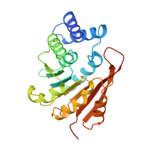
Crystal structure of catechol O-methyltransferase.

Vidgren, J.Svensson, L.A.Liljas, A.

(1994) Nature 368: 354-358

  • DOI: https://doi.org/10.1038/368354a0
  • Primary Citation of Related Structures:  
    1VID

  • PubMed Abstract: 

    Catechol O-methyltransferase (COMT, EC 2.1.1.6) is important in the central nervous system because it metabolizes catecholamine neurotransmitters such as dopamine. The enzyme catalyses the transfer of the methyl group from S-adenosyl-L-methionine (AdoMet) to one hydroxyl group of catechols. COMT also inactivates catechol-type compounds such as L-DOPA. With selective inhibitors of COMT in combination with L-DOPA, a new principle has been realized in the therapy of Parkinson's disease. Here we solve the atomic structure of COMT to 2.0 A resolution, which provides new insights into the mechanism of the methyl transfer reaction. The co-enzyme-binding domain is strikingly similar to that of an AdoMet-dependent DNA methylase, indicating that all AdoMet methylases may have a common structure.


  • Organizational Affiliation

    Orion Corporation, Espoo, Finland.


Macromolecules
Find similar proteins by:  (by identity cutoff)  |  3D Structure
Entity ID: 1
MoleculeChains Sequence LengthOrganismDetailsImage
CATECHOL O-METHYLTRANSFERASE221Rattus norvegicusMutation(s): 0 
EC: 2.1.1.6
UniProt
Find proteins for P22734 (Rattus norvegicus)
Explore P22734 
Go to UniProtKB:  P22734
Entity Groups  
Sequence Clusters30% Identity50% Identity70% Identity90% Identity95% Identity100% Identity
UniProt GroupP22734
Sequence Annotations
Expand
  • Reference Sequence
Small Molecules
Ligands 3 Unique
IDChains Name / Formula / InChI Key2D Diagram3D Interactions
SAM
Query on SAM

Download Ideal Coordinates CCD File 
C [auth A]S-ADENOSYLMETHIONINE
C15 H22 N6 O5 S
MEFKEPWMEQBLKI-FCKMPRQPSA-N
DNC
Query on DNC

Download Ideal Coordinates CCD File 
D [auth A]3,5-DINITROCATECHOL
C6 H4 N2 O6
VDCDWNDTNSWDFJ-UHFFFAOYSA-N
MG
Query on MG

Download Ideal Coordinates CCD File 
B [auth A]MAGNESIUM ION
Mg
JLVVSXFLKOJNIY-UHFFFAOYSA-N
Binding Affinity Annotations 
IDSourceBinding Affinity
DNC BindingDB:  1VID IC50: 12 (nM) from 1 assay(s)
Experimental Data & Validation

Experimental Data

  • Method: X-RAY DIFFRACTION
  • Resolution: 2.00 Å
  • R-Value Work: 0.194 
  • R-Value Observed: 0.194 
  • Space Group: P 32 2 1
Unit Cell:
Length ( Å )Angle ( ˚ )
a = 51.3α = 90
b = 51.3β = 90
c = 168.5γ = 120
Software Package:
Software NamePurpose
X-PLORmodel building
X-PLORrefinement
DENZOdata reduction
SCALEPACKdata scaling
X-PLORphasing

Structure Validation

View Full Validation Report


Entry History 

Deposition Data

Revision History  (Full details and data files)

  • Version 1.0: 1996-07-11
    Type: Initial release
  • Version 1.1: 2008-03-24
    Changes: Version format compliance
  • Version 1.2: 2011-07-13
    Changes: Version format compliance
  • Version 1.3: 2024-02-14
    Changes: Data collection, Database references, Derived calculations, Other