1UR9

Interactions of a family 18 chitinase with the designed inhibitor HM508, and its degradation product, chitobiono-delta-lactone


Experimental Data Snapshot

  • Method: X-RAY DIFFRACTION
  • Resolution: 1.80 Å
  • R-Value Free: 0.255 
  • R-Value Work: 0.208 
  • R-Value Observed: 0.208 

wwPDB Validation   3D Report Full Report


Ligand Structure Quality Assessment 


This is version 2.1 of the entry. See complete history


Literature

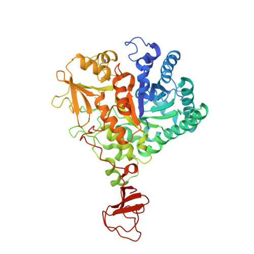
Interactions of a Family 18 Chitinase with the Designed Inhibitor Hm508 and its Degradation Product, Chitobiono-Delta-Lactone.

Vaaje-Kolstad, G.Vasella, A.Peter, M.G.Netter, C.Houston, D.R.Westereng, B.Synstad, B.Eijsink, V.G.H.Van Aalten, D.M.F.

(2004) J Biol Chem 279: 3612

  • DOI: https://doi.org/10.1074/jbc.M310057200
  • Primary Citation of Related Structures:  
    1UR8, 1UR9

  • PubMed Abstract: 

    We describe enzymological and structural analyses of the interaction between the family 18 chitinase ChiB from Serratia marcescens and the designed inhibitor N,N'-diacetylchitobionoxime-N-phenylcarbamate (HM508). HM508 acts as a competitive inhibitor of this enzyme with a K(i) in the 50 microM range. Active site mutants of ChiB show K(i) values ranging from 1 to 200 microM, providing insight into some of the interactions that determine inhibitor affinity. Interestingly, the wild type enzyme slowly degrades HM508, but the inhibitor is essentially stable in the presence of the moderately active D142N mutant of ChiB. The crystal structure of the D142N-HM508 complex revealed that the two sugar moieties bind to the -2 and -1 subsites, whereas the phenyl group interacts with aromatic side chains that line the +1 and +2 subsites. Enzymatic degradation of HM508, as well as a Trp --> Ala mutation in the +2 subsite of ChiB, led to reduced affinity for the inhibitor, showing that interactions between the phenyl group and the enzyme contribute to binding. Interestingly, a complex of enzymatically degraded HM508 with the wild type enzyme showed a chitobiono-delta-lactone bound in the -2 and -1 subsites, despite the fact that the equilibrium between the lactone and the hydroxy acid forms in solution lies far toward the latter. This shows that the active site preferentially binds the (4)E conformation of the -1 sugar, which resembles the proposed transition state of the reaction.


  • Organizational Affiliation

    Department of Chemistry and Biotechnology, Agricultural University of Norway, N-1432 As, Norway.


Macromolecules
Find similar proteins by:  (by identity cutoff)  |  3D Structure
Entity ID: 1
MoleculeChains Sequence LengthOrganismDetailsImage
CHITINASE B
A, B
499Serratia marcescensMutation(s): 1 
EC: 3.2.1.14
UniProt
Find proteins for Q54276 (Serratia marcescens)
Explore Q54276 
Go to UniProtKB:  Q54276
Entity Groups  
Sequence Clusters30% Identity50% Identity70% Identity90% Identity95% Identity100% Identity
UniProt GroupQ54276
Sequence Annotations
Expand
  • Reference Sequence
Oligosaccharides

Help

Entity ID: 2
MoleculeChains Length2D Diagram Glycosylation3D Interactions
2-acetamido-2-deoxy-beta-D-glucopyranose-(1-4)-2-(acetylamido)-2-deoxy-D-glucono-1,5-lactone
C, D
2N/A
Glycosylation Resources
GlyTouCan:  G08176ED
GlyCosmos:  G08176ED
Small Molecules
Ligands 3 Unique
IDChains Name / Formula / InChI Key2D Diagram3D Interactions
PHJ
Query on PHJ

Download Ideal Coordinates CCD File 
AC [auth B],
PA [auth A]
N-[(AMINOOXY)CARBONYL]-N-PHENYLAMINE
C7 H8 N2 O2
YQHOHPOCZUAUKP-UHFFFAOYSA-N
SO4
Query on SO4

Download Ideal Coordinates CCD File 
IA [auth A]
JA [auth A]
KA [auth A]
LA [auth A]
MA [auth A]
IA [auth A],
JA [auth A],
KA [auth A],
LA [auth A],
MA [auth A],
NA [auth A],
WB [auth B],
XB [auth B],
YB [auth B]
SULFATE ION
O4 S
QAOWNCQODCNURD-UHFFFAOYSA-L
GOL
Query on GOL

Download Ideal Coordinates CCD File 
AA [auth A]
AB [auth B]
BA [auth A]
BB [auth B]
CA [auth A]
AA [auth A],
AB [auth B],
BA [auth A],
BB [auth B],
CA [auth A],
CB [auth B],
DA [auth A],
DB [auth B],
E [auth A],
EA [auth A],
EB [auth B],
F [auth A],
FA [auth A],
FB [auth B],
G [auth A],
GA [auth A],
GB [auth B],
H [auth A],
HA [auth A],
HB [auth B],
I [auth A],
IB [auth B],
J [auth A],
JB [auth B],
K [auth A],
KB [auth B],
L [auth A],
LB [auth B],
M [auth A],
MB [auth B],
N [auth A],
NB [auth B],
O [auth A],
OA [auth A],
OB [auth B],
P [auth A],
PB [auth B],
Q [auth A],
QA [auth B],
QB [auth B],
R [auth A],
RA [auth B],
RB [auth B],
S [auth A],
SA [auth B],
SB [auth B],
T [auth A],
TA [auth B],
TB [auth B],
U [auth A],
UA [auth B],
UB [auth B],
V [auth A],
VA [auth B],
VB [auth B],
W [auth A],
WA [auth B],
X [auth A],
XA [auth B],
Y [auth A],
YA [auth B],
Z [auth A],
ZA [auth B],
ZB [auth B]
GLYCEROL
C3 H8 O3
PEDCQBHIVMGVHV-UHFFFAOYSA-N
Experimental Data & Validation

Experimental Data

  • Method: X-RAY DIFFRACTION
  • Resolution: 1.80 Å
  • R-Value Free: 0.255 
  • R-Value Work: 0.208 
  • R-Value Observed: 0.208 
  • Space Group: P 21 21 21
Unit Cell:
Length ( Å )Angle ( ˚ )
a = 55.524α = 90
b = 104.177β = 90
c = 185.992γ = 90
Software Package:
Software NamePurpose
CNSrefinement
DENZOdata reduction
SCALEPACKdata scaling
CNSphasing

Structure Validation

View Full Validation Report


Ligand Structure Quality Assessment 


Entry History 

Deposition Data

Revision History  (Full details and data files)

  • Version 1.0: 2004-04-27
    Type: Initial release
  • Version 1.1: 2015-04-15
    Changes: Database references, Derived calculations, Non-polymer description, Other, Refinement description, Source and taxonomy, Structure summary, Version format compliance
  • Version 2.0: 2020-07-29
    Type: Remediation
    Reason: Carbohydrate remediation
    Changes: Atomic model, Data collection, Derived calculations, Other, Structure summary
  • Version 2.1: 2023-12-13
    Changes: Data collection, Database references, Refinement description, Structure summary