1U4J

Crystal structure of a carbohydrate induced dimer of group I phospholipase A2 from Bungarus caeruleus at 2.1 A resolution


Experimental Data Snapshot

  • Method: X-RAY DIFFRACTION
  • Resolution: 2.18 Å
  • R-Value Free: 0.220 
  • R-Value Work: 0.194 
  • R-Value Observed: 0.194 

wwPDB Validation   3D Report Full Report


This is version 1.5 of the entry. See complete history


Literature

Crystal structure of a carbohydrate induced homodimer of phospholipase A(2) from Bungarus caeruleus at 2.1A resolution

Singh, G.Gourinath, S.Sarvanan, K.Sharma, S.Bhanumathi, S.Betzel, C.Yadav, S.Srinivasan, A.Singh, T.P.

(2005) J Struct Biol 149: 264-272

  • DOI: https://doi.org/10.1016/j.jsb.2004.11.011
  • Primary Citation of Related Structures:  
    1U4J

  • PubMed Abstract: 

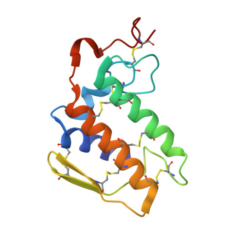
    This is the first crystal structure of a carbohydrate induced dimer of phospholipase A(2) (PLA(2)). This is an endogenous complex formed between two PLA(2) molecules and two mannoses. It was isolated from Krait venom (Bungarus caeruleus) and crystallized as such. The complete amino acid sequence of PLA(2) was determined using cDNA method. Three-dimensional structure of the complex has been solved with molecular replacement method and refined to a final R-factor of 0.192 for all the data in the resolution range 20.0-2.1A. The presence of mannose molecules in the protein crystals was confirmed using dinitrosalicylic acid test and the molecular weight of the dimer was verified with MALDI-TOF. As indicated by dynamic light scattering and analytical ultracentrifugation the dimer was also stable in solution. The good quality non-protein electron density at the interface of two PLA(2) molecules enabled us to model two mannoses. The mannoses are involved extensively in interactions with protein atoms of both PLA(2) molecules. Some of the critical amino acid residues such as Asp 49 and Tyr 31, which are part of the substrate-binding site, are found facing the interface and interacting with mannoses. The structure of the complex clearly shows that the dimerization is caused by mannoses and it results in the loss of enzymatic activity.


  • Organizational Affiliation

    Department of Biophysics, All India Institute of Medical Sciences, New Delhi 110 029, India.


Macromolecules
Find similar proteins by:  (by identity cutoff)  |  3D Structure
Entity ID: 1
MoleculeChains Sequence LengthOrganismDetailsImage
phospholipase A2 isoform 2
A, B
118Bungarus caeruleusMutation(s): 0 
EC: 3.1.1.4
Membrane Entity: Yes 
UniProt
Find proteins for Q6SLM1 (Bungarus caeruleus)
Explore Q6SLM1 
Go to UniProtKB:  Q6SLM1
Entity Groups  
Sequence Clusters30% Identity50% Identity70% Identity90% Identity95% Identity100% Identity
UniProt GroupQ6SLM1
Sequence Annotations
Expand
  • Reference Sequence
Experimental Data & Validation

Experimental Data

  • Method: X-RAY DIFFRACTION
  • Resolution: 2.18 Å
  • R-Value Free: 0.220 
  • R-Value Work: 0.194 
  • R-Value Observed: 0.194 
  • Space Group: H 3
Unit Cell:
Length ( Å )Angle ( ˚ )
a = 80.36α = 90
b = 80.36β = 90
c = 99.44γ = 120
Software Package:
Software NamePurpose
CNSrefinement
MAR345data collection
SCALEPACKdata scaling
AMoREphasing

Structure Validation

View Full Validation Report


Entry History 

Deposition Data

Revision History  (Full details and data files)

  • Version 1.0: 2004-08-10
    Type: Initial release
  • Version 1.1: 2008-04-30
    Changes: Version format compliance
  • Version 1.2: 2011-07-13
    Changes: Version format compliance
  • Version 1.3: 2017-10-11
    Changes: Refinement description
  • Version 1.4: 2020-07-29
    Type: Remediation
    Reason: Carbohydrate remediation
    Changes: Advisory, Data collection, Derived calculations, Structure summary
  • Version 1.5: 2023-10-25
    Changes: Data collection, Database references, Refinement description, Structure summary