1TB3

Crystal Structure Analysis of Recombinant Rat Kidney Long-chain Hydroxy Acid Oxidase


Experimental Data Snapshot

  • Method: X-RAY DIFFRACTION
  • Resolution: 2.30 Å
  • R-Value Free: 0.269 
  • R-Value Work: 0.239 

wwPDB Validation   3D Report Full Report


Ligand Structure Quality Assessment 


This is version 1.3 of the entry. See complete history


Literature

Crystal Structure Analysis of Recombinant Rat Kidney Long Chain Hydroxy Acid Oxidase.

Cunane, L.M.Barton, J.D.Chen, Z.W.Le, K.H.D.Amar, D.Lederer, F.Mathews, F.S.

(2005) Biochemistry 44: 1521-1531

  • DOI: https://doi.org/10.1021/bi048616e
  • Primary Citation of Related Structures:  
    1TB3

  • PubMed Abstract: 

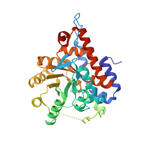
    Long chain hydroxy acid oxidase (LCHAO) is a member of an FMN-dependent enzyme family that oxidizes L-2-hydroxy acids to ketoacids. LCHAO is a peroxisomal enzyme, and the identity of its physiological substrate is unclear. Mandelate is the most efficient substrate known and is commonly used in the test tube. LCHAO differs from most family members in that one of the otherwise invariant active site residues is a phenylalanine (Phe23) instead of a tyrosine. We now report the crystal structure of LCHAO. It shows the same beta8alpha8 TIM barrel structure as other structurally characterized family members, e.g., spinach glycolate oxidase (GOX) and the electron transferases yeast flavocytochrome b2 (FCB2) and Pseudomonas putida mandelate dehydrogenase (MDH). Loop 4, which is mobile in other family members, is visible in part. An acetate ion is present in the active site. The flavin interacts with the protein in the same way as in the electron transferases, and not as in GOX, an unexpected observation. An interpretation is proposed to explain this difference between GOX on one hand and FCB2 and LCHAO on the other hand, which had been proposed to arise from the differences between family members in their reactivity with oxygen. A comparison of models of the substrate bound to various published structures suggests that the very different reactivity with mandelate of LCHAO, GOX, FCB2, and MDH cannot be rationalized by a hydride transfer mechanism.


  • Organizational Affiliation

    Department of Biochemistry and Molecular Biophysics, Washington University School of Medicine, St. Louis, Missouri 63110, USA.


Macromolecules
Find similar proteins by:  (by identity cutoff)  |  3D Structure
Entity ID: 1
MoleculeChains Sequence LengthOrganismDetailsImage
Hydroxyacid oxidase 3
A, B, C, D, E
A, B, C, D, E, F, G, H
352Rattus norvegicusMutation(s): 0 
Gene Names: HAO3HAOX3
EC: 1.1.3.15
UniProt
Find proteins for Q07523 (Rattus norvegicus)
Explore Q07523 
Go to UniProtKB:  Q07523
Entity Groups  
Sequence Clusters30% Identity50% Identity70% Identity90% Identity95% Identity100% Identity
UniProt GroupQ07523
Sequence Annotations
Expand
  • Reference Sequence
Small Molecules
Ligands 2 Unique
IDChains Name / Formula / InChI Key2D Diagram3D Interactions
FMN
Query on FMN

Download Ideal Coordinates CCD File 
I [auth A]
K [auth B]
M [auth C]
O [auth D]
Q [auth E]
I [auth A],
K [auth B],
M [auth C],
O [auth D],
Q [auth E],
S [auth F],
U [auth G],
W [auth H]
FLAVIN MONONUCLEOTIDE
C17 H21 N4 O9 P
FVTCRASFADXXNN-SCRDCRAPSA-N
ACY
Query on ACY

Download Ideal Coordinates CCD File 
J [auth A]
L [auth B]
N [auth C]
P [auth D]
R [auth E]
J [auth A],
L [auth B],
N [auth C],
P [auth D],
R [auth E],
T [auth F],
V [auth G],
X [auth H]
ACETIC ACID
C2 H4 O2
QTBSBXVTEAMEQO-UHFFFAOYSA-N
Experimental Data & Validation

Experimental Data

  • Method: X-RAY DIFFRACTION
  • Resolution: 2.30 Å
  • R-Value Free: 0.269 
  • R-Value Work: 0.239 
  • Space Group: P 21 21 21
Unit Cell:
Length ( Å )Angle ( ˚ )
a = 114.839α = 90
b = 151.1β = 90
c = 222.36γ = 90
Software Package:
Software NamePurpose
HKL-2000data collection
SCALEPACKdata scaling
AMoREphasing
CNSrefinement
HKL-2000data reduction

Structure Validation

View Full Validation Report


Ligand Structure Quality Assessment 


Entry History 

Deposition Data

Revision History  (Full details and data files)

  • Version 1.0: 2005-02-01
    Type: Initial release
  • Version 1.1: 2008-04-30
    Changes: Version format compliance
  • Version 1.2: 2011-07-13
    Changes: Version format compliance
  • Version 1.3: 2023-08-23
    Changes: Data collection, Database references, Derived calculations, Refinement description