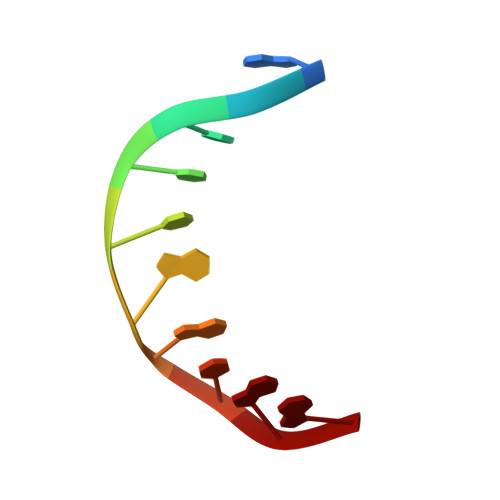
Effect of bulky lesions on DNA: Solution structure of a DNA duplex containing a cholesterol adduct.
Gomez-Pinto, I., Cubero, E., Kalko, S.G., Monaco, V., Van Der Marel, G., Van Boom, J.H., Orozco, M., Gonzalez, C.(2004) J Biol Chem 279: 24552-24560
- PubMed: 15047709
- DOI: https://doi.org/10.1074/jbc.M311751200
- Primary Citation of Related Structures:
1SP6, 1SSJ - PubMed Abstract:
The three-dimensional solution structure of two DNA decamers of sequence d(CCACXGGAAC)-(GTTCCGGTGG) with a modified nucleotide containing a cholesterol derivative (X) in its C1 '(chol)alpha or C1 '(chol)beta diastereoisomer form has been determined by using NMR and restrained molecular dynamics. This DNA derivative is recognized with high efficiency by the UvrB protein, which is part of the bacterial nucleotide excision repair, and the alpha anomer is repaired more efficiently than the beta one. The structures of the two decamers have been determined from accurate distance constraints obtained from a complete relaxation matrix analysis of the NOE intensities and torsion angle constraints derived from J-coupling constants. The structures have been refined with molecular dynamics methods, including explicit solvent and applying the particle mesh Ewald method to properly evaluate the long range electrostatic interactions. These calculations converge to well defined structures whose conformation is intermediate between the A- and B-DNA families as judged by the root mean square deviation but with sugar puckerings and groove shapes corresponding to a distorted B-conformation. Both duplex adducts exhibit intercalation of the cholesterol group from the major groove of the helix and displacement of the guanine base opposite the modified nucleotide. Based on these structures and molecular dynamics calculations, we propose a tentative model for the recognition of damaged DNA substrates by the UvrB protein.
Organizational Affiliation:
Instituto de Química Física Rocasolano, Consejo Superior de Investigaciones Científicas, C/. Serrano 119, 28006 Madrid, Spain.